
We are delighted to announce the 71st meeting of the Danish Society for Flow Cytometry (DSFCM), focusing on Cell Sorting.
The event will be held at:
Program Outline: (for more details see program)
About the event:
Registration:
Follow the link to register to the event ! https://events.au.dk/71stdsfcm

Registration deadline October 31st 2024 !
The 70th meeting and the annual general assembly in DSFCM will take place on April 15, 2024, from 11 AM – 7 PM at DTU,
DTU Lyngby campus
Anker Engelunds Vej 1
Bygning 101A, 1st floor, meeting room M1
2800 Kgs. Lyngby
Program outline (for more details see program)
11 AM – 4 PM Join us for an interesting program with the subject Flow Cytometry in Immunology
4 – 5 PM General assembly
5 – 7 PM Join us for light dinner, a drink, and networking (members only)
Registration https://events.au.dk/70thdsfcm
Registration deadline: April 1st

Incoming suggestions for the general assembly must be sent to Anja B Bohn at anja@biomed.au.dk the latest 2 PM April 12th
The theme for the meeting is “Panel Design and Gating”, where we have invited people to come and share their knowledge in this field.
Program for the meeting.
Meeting is: November 9th 2023, kl 11:00-18:00, Eduard Biermann auditorium (1252-204), University of Aarhus, “Søauditorierne”,
Bartholins Allé 3. DK-8000 Aarhus C
Registration deadline October 25th 2023
The annual meeting in the society will be May 3rd in Copenhagen. We start with General assembly and then proceed to the program with the theme “Functional Flow Cytometry Assays”. The evening ends with a light dinners for members of the society. If you are interested in becoming a member click here.
Program and registration is now open. Last change for registration is 19th April 2023.
Map of the venue, it is in 13A the auditorium is located.
The Autumn meeting in DSFCM will be November 3rd at Moesgård Museum https://www.moesgaardmuseum.dk/. We offer a short guided tour on the museum before we start the meeting. The program will be about spectral flow cytometry, we have invited several speakers to cover the subject, so save the date.
Please find program and link for signing up at the meeting.
Link for participation: https://events.au.dk/67thdsfcm/conference
Deadline for participating is October 25 2022.
It is our pleasure to invite you to the 66th meeting of the Danish society for flow cytometry.
The theme for this meeting will be “Challenges in Flow Cytometry“.
Keynote speaker is Andrew Filby, Faculty of Medical Sciences, Newcastle University, UK.
At this meeting we encourage students and researchers to share problems, challenges and troubleshooting related to flow cytometry and imaging flow cytometry. When signing up, choose if you would like to give an oral presentation or present a poster. We anticipate the oral presentation to be 15min each
Students giving an oral presentation at this meeting, will receive a one-year free membership of the Danish Society for Flow Cytometry.
The program can be found here, remember that you need to sign up for the meeting so we can buy coffee, dinner etc, registration up at https://events.au.dk/66thdsfcm
The evening program (from 18:00-20:00) is only for members of the society – If you are not a member yet, it only takes a few minutes to become one to attend the social event: https://flowcytometri.dk/join-dsfcm/
We continue to have our meetings online. We have now prepared “Exotic Flow Cytometry Vol 2”.
It is our pleasure to invite you to the 65th meeting in DSFCM. The topic for this meeting will be “Exotic Flow Cytometry”, followed by general assembly.
You can see the full program here.
13.00-13.10 Welcome
13.10-13.55 Jaroslav Dolezel, Prof., Institute of Experimental Botany; Olomouc; Czech Republic. “Flow cytogenetics: the exotic way of genome sequencing, gene cloning and uncovering chromosome ultrastructure” See presentation
13.55-14.05 Break
14.05-14.25 Tina Santl-Temkiv, Asst.Prof. Department of Biology, Aarhus. “Sea-spray experiments coupled with flow cytometry reveal preferential emissions of microorganisms from aqueous environments into the atmosphere”
14.30-14.50 Kasper Urup Kjeldsen, Assoc.Prof, Department of Biology, Aarhus “Fluorescence-activated cell sorting for studying microbial dark matter”
14.50-15.00 Break
15.00-16.00 General assembly
Taking the current situation in Denmark we have a decided to hold our next meeting as a web meeting.
It is our pleasure to invite you to the 64th meeting in DSFCM. The topic for this meeting will be “Exotic Flow Cytometry and SARS-CoV-2 in flow cytometry laboratories”.
You can see the full program here.
13.00-13.10 Welcome
13.10-13.40 Samplix “Double-emulsion droplet sorting: Strategies and applications for multiomic profiling”, see the presentation.
13.45-14.15 VikingGenetics “Through the years from sperm sorting, to sex-sorting and QC”, see the presentation.
14.20-14.50 How are we dealing with Corona in flow cytometry laboratories – a short introduction by Jan Pravsgaard followed by a discussion, see the presentation.
14.50-15.00 Break
15.00-16.00 General assembly
The program for the 62nd meeting of DSFCM is now ready. The meeting will be held at Aarhus University Hospital, Aud G, Entrance G, November 12th 2019
Palle Juul-Jensens Boulevard 99
8200 Aarhus N
Danmark
Map of venue: https://www.auh.dk/siteassets/patient/find-vej/auh-oversigtskort-030619.pdf
The theme of the upcoming meeting is “Flow Cytometry in Clinical Research”.
Program and registration. Abstract for the presentations.
Please not, you need to sign up for the meeting so we can order coffee and lunch.
The program for the 61st meeting of DSFCM is now ready. The meeting will be held at Department of Health Technology – DTU Health Tech, May 27th 2019.
Anker Engelundsvej 1, Building 101A
2800 Kgs Lyngby
Danmark
DTU Meeting Center, 1st floor, Meeting room 1
https://www.dtu.dk/english/about/campuses/dtu-lyngby-campus/getting-there
The theme of the upcoming meeting is “Detection of extracellular vesicles in flow cytometry”. Four great speakers have confirmed their attendance:
Program and registration. Abstract for the presentations.
Please not, you need to sign up for the meeting so we can order coffee and lunch.
60th Meeting of DSFCM
Thursday, 1 November 2018, 11:00-19:00.
Location: Holst auditorium, The Maersk Tower, Blegdamsvej 3B, 2200 København N
Program
11:00-17:00 Welcome
The meeting is open for everyone interested.
Please register via this link: https://events.au.dk/DSFCM60Meetingand30anniversary/sign%2Dup.html
59th Meeting of DSFCM
Tuesday, 17 April 2018, 11:00-16:00.
Naturhistorisk Museum, Aarhus University, Wilhelm Meyers Allé 10, DK-8000 Aarhus (map).
Final program
All are welcome. Attendance is free of charge.
Please, register via email to Charlotte Christie Petersen at ccp@biomed.au.dk with ”59th DSFCM meeting” as headline no later than Wednesday, 11 April 2018.
Rev 3 June 2018 /JKL
58th Meeting of DSFCM
Nordic Flow Cytometry Meeting
(58th Meeting of DSFCM)
DGI-byen, Tietgensgade 65, 1704 Copenhagen V, Denmark.
August 30 – September 1, 2017.
Program and Abstract Book
Registration here.
Abstract submission here.
Signup for course at University of Copenhagen here.
Exhibition registration here.
Sponsor registration here.

Rev 30 June 2017 /JKL
57th Meeting of DSFCM
Agilent Technologies Denmark ApS, Produktionsvej 42, 2600 Glostrup.
28 March 2017, 10:00-16:00.
Registration is mandatory. Please register via email to jpc@sund.ku.dk with ”57th dsfcm meeting” as headline no later than Tuesday 07.03.2017; include your full name; include if you wish to participate in tour on Agilent.
Rev 24 April 2017 /JKL
56th Meeting of DSFCM
Auditorium 1, Aarhus Universitetshospital, Tage-Hansens Gade 2, Indgang 4A, 8000 Aarhus C (map).
3 November 2016, 11:00-16:00.
New Applications and Protocols in Flow Cytometry (program)
Please register via email no later than 21 October 2016 (see program). <
Rev 6 December 2016 /JKL
55th Meeting of DSFCM
5. April 2016, kl 10:00-16:00.
Auditorium, Entrance 24 (next to staff cafeteria), Roskilde Hospital, Køgevej 7-13, 4000 Roskilde (Map).
Rev 15 April 2016 /JKL
54th Meeting of DSFCM
October 29, 2015, 11:15-15:45.
Auditorium at Naturhistorisk Museum, Wilhelm Meyers Allé 210, Aarhus University, DK-8000 Aarhus C (map).
Safety in flow cytometry (program):
All are welcome. For registration, see program.
Rev 10 November 2015 /JKL
Please register via email to Jan P. Christensen with “attending at 53th meeting” as headline no later than Tuesday 17 March 2015.
Rev 31 March 2015 /JKL
52nd Meeting of DSFCM
May 6, 2014, 11:00-15:45.
Auditorium 1, Rigshospitalet, Entrance 44, Blegdamsvej 9, 2100 København Ø (map).
All are welcome and attendance is free of charge, however, registration is required. Please register via email to jpc@sund.ku.dk with ”52nd dsfcm meeting” as headline no later than Friday 25.04.2014.
For travel grants and commercial exhibition, see Program.
50th Meeting of DSFCM:
4 April 2013, 11:00-16:30.
Auditorium 93, Rigshospitalet, Entrance 93, Juliane Maries Vej, DK-2100 Copenhagen Ø (vejkort).
Microorganisms and flow cytometry
Keynote lecture
Plenary talks
Registration: All are wellcome, but registration is necessary because of limited number of seats. No registration fee. Please, register by e-mail to dsfcm.dk@gmail.com, subject “50th DSFCM Meeting”, no later than 22 March 2013.
Exhibition: For corporate members of DSFCM, sales pitches of 1 x 1.5 m are available. Please, register by e-mail to dsfcm.dk@gmail.com, subject “stand at 50th meeting”, no later than 22 March 2013.
Generalforsamling 2013 afholdes i tilknytning til det videnskabelige møde.
Rev 10 April 2013 /JKL
49th Meeting of the Danish Society for Flow Cytometry.
25 October 2012, 15:00-17:00, Haderup Auditorium, Panum Institute, Nørre Allé 20, Copenhagen N.
Progress in Flow Cytometry
Program:
All are welcome. No registration fee, but registration is necessary.
Register to the organizer, Jacob Larsen, by mail to jacl@regionsjaelland.dk.
Rev 2 November 2012 /JKL
48th Meeting of the Danish Society for Flow Cytometry.
28 March 2012, 11:00–16:00. ALK-Abelló, Bøge Alle 6-8, Hørsholm.
All are wellcome, but registration is necessary because of limited number of seats. No registration fee. Free lunch. Register by e-mail to Alexander Schmitz (alex.schmitz@rn.dk), subject: 48th Meeting.
The meeting had 53 participants (registration closed at this number)
I. Generalforsamling for DSFCM
.
II. New faces & Instruments in Danish Flow Cytometry 2012
Rev 24 April 2012 /JKL
47th Meeting of the Danish Society for Flow Cytometry.
Program (oversigt)
Tilføjelse til programmet:
Foredrag af Christiane Beer om FCM af micronuclei.
Rev 20 Oct 2011 /JKL
46th Meeting of the Danish Society for Flow Cytometry.
![]()
![]()
![]()
SCANDINAVIAN FLOW CYTOMETRY MEETING, Halmstad, Sweden, April 4 (11:00) – April 5 (15:00), 2011.
Flow cytometry in research and clinical practice.
To members of the Danish Society for Flow Cytometry and others interested!
It is a pleasure to announce that the 46th meeting of DSFCM will take place as a joint meeting with Svensk Förening för Flödescytometri (SFFF) and Norsk Flowcytometriforening (NFCF). We hope for a wide participation of Danish users of cytometry from the clinic, research and industry as well as corporate participation from producers and traders of cytometers and reagents.
Program (revised 22 March 2011).
The program includes plenary lectures by:
The program also includes a poster session and workshops organized by companies.
Participants are encouraged to submit ABSTRACTS for the sessions of posters and free lectures (deadline for submission to Tom Lundahl was February 28).
Registration form (doc, pdf). Participation costs 1500 SEK for both days. Lodging has to be booked independently; see suggestions for hotels and travel routes in the registration form.
The language of the meeting is English or Scandinavian, according to language of session speakers.
The local meeting organizer is: Tom Lundahl. Representatives of DSFCM in the program comittee are: Carl-Henrik Brogren og Jacob Larsen.
SKANDINAVISK FLOWCYTOMETRI MØDE, Halmstad, Sverige, 4. april (11:00) – 5. april (15:00) 2011.
Flowcytometri i klinikkens og forskningens tjeneste.
Til medlemmerne af Dansk Selskab for Flowcytometri og alle interesserede!
Det er en glæde at meddele, at det næste årsmøde i DSFCM (DSFCM’s 46. møde) vil blive afholdt i fællesskab med Svensk Förening för Flödescytometri (SFFF) og Norsk Flowcytometriforening (NFCF). Vi håber på en bred deltagelse af danske cytometribrugere fra klinikken, forskningen og industrien såvel som deltagelse af firmaerne for cytometre og reagenser.
Program (revideret 22.3.2011).
Programmet indeholder plenarforedrag af:
Programmet indeholder ud over sessioner med foredrag også en poster session samt workshops arrangerede af firmaer.
Deltagere opfordres til at indsende ABSTRACTS til sessionerne med postere og frie foredrag (tidsfrist for indsendelse til Tom Lundahl var 28. februar) .
Tilmeldingsformular (doc, pdf). Deltagelse i mødet koster 1500 SEK for begge dage. Logi skal bestilles særskilt; se forslag til hoteller og rejseruter i tilmeldingsformularen.
Mødets sprog er engelsk eller skandinaviske sprog, afhængigt af den enkelte sessions talere.
Lokal mødearrangør er: Tom Lundahl. DSFCM’s bestyrelses repræsentanter i programkomitéen er: Carl-Henrik Brogren og Jacob Larsen.
45th Meeting of the Danish Society for Flow Cytometry
Flowcytometri og fertilitet
(human fertilitet, kønsselektion i veterinærindustrien, etiske aspekter).
Mødearrangør: Jens Peter Stenvang. Abstracts (PDF).
Følgende bestyrelsesmedlemmer er fratrådt eller ønsker af fratræde:
Til supplering af Bestyrelsen (1 medlem) og suppleanter (2 medlemmer) peges på følgende DSFCM-medlemmer, der har erklæret sig som kandidater ved valget:
Bestyrelsen afholdt den 22-23. januar 2010 et internatmøde i et tilsneet sommerhus i Højby ved Sejerø Bugt, hvor der blev diskuteret og drøftet emner inden for flow cytometri og forskning i relation til mulige fremtidige aktiviteter for DSFCM.
Til stede var Hans Jürgen Hoffmann, Alexander Schmitz, Carl-Henrik Brogren, Thomas Hviid og Jørgen K. Larsen. Hele bestyrelsen bortset fra Jens Peter stenvang, som havde måttet melde afbud, var således til stede. Endvidere deltog to kandidater til det kommende bestyrelsesvalg, Jakob Larsen og Rasmus Foldbjerg. En tredie kandidat, Mikkel Steen Petersen, kunne ikke deltage på grund af andre aftaler og kort varsel.
Blandt emner, der vil blive drøftet i formandens beretning til generalforsamlingen og under kommende møder, kan nævnes følgende:
På bestyrelsens vegne,
Hans Jürgen Hoffmann, formand
44. Møde i Dansk Selskab for Flowcytometri
Fredag, 29. maj 2009, Foredragssalen, Roskilde Sygehus, Roskilde.
Alle er velkomne. Tilmelding ikke nødvendig.
A) Leukæmi- og lymfomdiagnostik med flowcytometri.
Mødearrangør: Thomas Hviid.
B) Generalforsamling i Dansk Selskab for Flowcytometri.
9 June 2009 /JKL
43rd Meeting of the Danish Society for Flow Cytometry
Flowcytometri-workshop – data management og kvalitetssikring
28. august 2008, 09.00-12.15. Odense Congress Center, Odense.
DEKS brugermøde 2008: Workshop I, Lokale 24.
Mødearrangør og mødeleder: Hans Jürgen Hoffmann.
Tilmelding er nødvendig. Medlemmer af DSFCM kan deltage gratis i workshoppen inklusive kaffe/kage og frokost, hvis de tilmelder sig ved e-mail til Hans Jürgen Hoffmann senest 25. juli, hvorimod ikke-medlemmer af DSFCM må foretage normal tilmelding tilDEKS brugermøde 2008.
42nd Meeting of the Danish Society for Flow Cytometry
Monday, 5 May 2008
Organizers:
Carl-Henrik Brogen, Dept. of Medical Biochemistry and Genetics, Faculty of Health Sciences, University of Copenhagen, DK.
Graham Leslie, Inst. of Medical Biology – Immunology and Microbiology, University of Southern Denmark, Odense, DK.
Hans Jürgen Hoffmann, Dept. of Respiratory Diseases, Inst. of Clinical Medicine, Aarhus University Hospital, Aarhus, DK.
Program
Dam Auditorium, Panum Building
10:00-12:00. Session 1:
20 years of flow cytometry with the DSFCM
Chair: Hans Jürgen Hoffmann and Carl-Henrik Brogren.
12:00-13:00
Posters viewing and Corporate Exhibitions with a buffet lunch
13:00-15:00. Session 2:
Flow Cytometry Core Facilities
Chair: Graham Leslie, and Alexander Schmitz, Inst. of Molecular Biology, Aarhus University, Aarhus, DK.
15:00-15:40. Parallel A
Posters viewing and Corporate Exhibitions with a coffee break
15:10-15:40. Parallel B
Generalforsamling (Annual General Meeting of DSFCM, in Danish)
*
**
15:40-16:30
Panel discussion on Flow Cytometry Core Facilities
Participants: Derek Davies, Willem E. Corver, Andrew Riddell, Jan Grawe, Alexander Schmitz, Lars Ryder (Department of Clinical Immunology, Rigshospitalet, Copenhagen, DK), and Graham Leslie (Chairman).
16:30-17:00
Poster presentations
Chair: Carl-Henrik Brogren.
17:00-17:30
Reception with election of the best poster
17:30-
Dinner Party
Registration
dinner party
Posters
Exhibition by corporate members
Revised, 4 May 2008 /JKL /www.flowcytometry.dk
41st Meeting of the Danish Society for Flow Cytometry.
Sarcoidosis: clinical & immunological features and ex vivo diagnosis
October 3, 2007, 17:00-20:00, Auditorium 3, Building 1252 (Søauditorierne), Wilhelm Meyers Allé, Aarhus University, 8000 Aarhus C.
The meeting is organized by the Danish Societies for Flow Cytometry, Respiratory Medicine and Rheumatology, and is partially funded by Schering-Plough – Organizer, DSFCM: Hans Jürgen Hoffmann.
4 Oct 2007 /JKL
40th Meeting of the Danish Society for Flow Cytometry.
September 20, 2007, 13:20–16:15, Room 7+8, Odense Congress Center, Odense.
Workshop on Quality Assurance for Flow Cytometry under the auspicies of DEKS
Program, see DEKS Brugermøde 2007 (Workshop II: Kvalitetssikring af flowcytometri).
Organizer: Hans Jürgen Hoffmann. Chair: Tom Just.
Register by mail to Hans Jürgen Hoffmann before 13 september 2007, with indication whether you join us for lunch (buffet is arranged by DEKS in Sal Jylland at 12:15-13:15). Participation in the workshop, including lunch, is free for members of DSFCM. Registration fee for other participants to be announced by DEKS.
21 Sept 2007 /JKL
39th Meeting of the Danish Society for Flow Cytometry
Joint meeting of the Danish Society for Cyto- and Histochemistry and DSFCM.
Advanced fluorescence imaging techniques
Time Wednesday, 7 March 2007, 16:00-19:00.
Location Auditorium 1-01 (Festauditoriet), Faculty of Life Sciences. University of Copenhagen, Frederiksberg Campus, Bülowsvej 17, 1870 Frederiksberg C, Denmark.
Registration Until 2 March 2007 to Ulla Evald, Institute of Pathology, BBH, at ue01@bbh.hosp.dk.
All are welcome.
Program Chair: Lars-Inge Larsson.
16.00–16.05 Introduction by Hans Lyon and Lars-Inge Larsson.
16.05–16.35 Bo van Deurs, Department of Cellular and Molecular Medicine, the Panum Building, University of Copenhagen:
FRAP determination of caveolar mobility in relation to EGFR endocytosis.
16.35–17.05 Claudia Lukas, Institute of Cancer Biology and Centre for Genotoxic Stress Research, Danish Cancer Society, Strandboulevarden 49, DK-2100 Copenhagen, Denmark:
DNA damage-induced cell cycle checkpoints and their dynamics in living mammalian cells.
17.05–17.35 Alexander Schulz, Plant Physiology and Anatomy Laboratory, Department of Plant Biology, Faculty of Life Sciences. University of Copenhagen, Frederiksberg Campus:
Bioimaging with ”caged probes”.
17.35–18.05 Christoffer Lagerholm, MEMPHYS – Center for Biomembrane Physics, University of Southern Denmark:
Biological Applications of Quantum Dots.
18.05–18.30 Discussion by the panel of speakers.
18.30–19.00 Refreshments.
Abstracts
FRAP determination of caveolar mobility in relation to EGFR endocytosis
Bo van Deurs, Department of Cellular and Molecular Medicine, the Panum Building, University of Copenhagen.
It is well-established that following Epidermal Growth Factor (EGF) binding the EGF receptor (EGFR) becomes internalized by clathrin-dependent endocytosis. However, recently it was reported that caveolae-dependent endocytosis is involved in the uptake of EGFR at high concentrations of ligand. We have previously shown that plasma membrane caveolae are stable membrane domains not involved in constitutive endocytosis [1]. We therefore investigated whether stimulation with high concentrations (100 ng/ml) of EGF induced mobilization of plasma membrane caveolae, either as a bulk movement of cell surface caveolae towards the interior of the cell, or as an increased turnover of caveolae at the plasma membrane [2]. Live-cell microscopy of cells expressing GFP-Caveolin-1 as a marker for caveolae revealed that no net movement of caveolae takes place in cells stimulated with high concentrations of EGF. In addition, Fluorescence Recovery after Photobleaching (FRAP) analysis of GFP-labeled plasma membrane caveolae showed that EGF stimulation does not increase the turnover of caveolae at the plasma membrane. Both in control cells and in EGF stimulated cells, the mobile fraction of caveolae was as low as 20-30%. In contrast, we found that endocytosis of EGFR was efficiently inhibited by knockdown of clathrin heavy chain, both at high and low concentrations of EGF [2]. Our results show that caveolae are not involved in endocytosis of EGF-bound EGFR to any significant degree, and high concentrations of EGF do not mobilize caveolae.
References:
[1] Thomsen, P., K.Roepstorff, M.Stahlhut, and B.van Deurs. Caveolae are highly immobile plasma membrane microdomains, which are not involved in constitutive endocytic trafficking. Mol. Biol. Cell 13, 238-250 (2002).
[2] Kazazic, M., K.Roepstorff, L.E.Johannessen, N.M.Pedersen, B.van Deurs, E.Stang, and I.H.Madshus. EGF-induced activation of the EGF receptor does not trigger mobilization of caveolae. Traffic. 7, 1518-1527 (2006).
DNA damage-induced cell cycle checkpoints and their dynamics in living mammalian cells
Claudia Lukas, Institute of Cancer Biology and Centre for Genotoxic Stress Research, Danish Cancer Society, Strandboulevarden 49, DK-2100 Copenhagen, Denmark (clukas@cancer.dk).
To protect the genome against adverse effects of DNA damage, eukaryotic cells evolved surveillance pathways, so-called ‘checkpoints’ that delay cell cycle progression until the productive repair of the primary DNA lesions. Our laboratory is interested in how the key checkpoint-associated molecular network operates in its physiological environment, the nucleus of a living mammalian cell. I will discuss the current advances in real-time imaging of molecular trafficking inside the nucleus and provide evidence that interaction of distinct checkpoint complexes with the sites of DNA damage is tightly regulated in space and time. I will show that the mode of protein redistribution in and out of the damaged nuclear compartments might lead to discoveries of new functions of these factors, and thus provide a more complete picture of the basic principles of functional connection between the spatially restricted sites of DNA lesions and the pan-nuclear cell cycle effectors.
References:
1) Lukas, C., Falck, J., Bartkova, J., Bartek, J., and Lukas, J. Distinct spatio-temporal dynamics of mammalian checkpoint regulators induced by DNA damage. Nat. Cell Biol. 5, 255-260 (2003).
2) Lukas, C., Melander, F., Stucki, M., Falck, J., Bekker-Jensen, S., Goldberg, M., Lerenthal, Y., Jackson, S. P., Bartek, J., and Lukas, J. Mdc1 couples DNA double-strand break recognition by Nbs1 with its H2AX-dependent chromatin retention. EMBO J. 23, 2674-2683 (2004).
3) Lukas, J., Lukas, C., and Bartek, J. Mammalian cell cycle checkpoints: Signalling pathways and their organization in space and time. DNA Repair, 3, 997-1007 (2004).
4) Bekker-Jensen, S., Lukas, C., Melander, F., Bartek, J., and Lukas, J. Dynamic assembly and sustained retention of 53BP1 at the sites of DNA damage are controlled by Mdc1/NFBD1. J Cell Biol. (2), 201-211 (2005).
5) Bekker-Jensen S., Lukas, C., Kitagawa R., Kastan, M.B., Bartek, J., and Lukas, J. Spatial organization of the mammalian genome surveillance machinery in response to DNA strand breaks. J Cell Biol.(2), 195-206 (2006).
Bioimaging with ”caged probes”
Alexander Schulz, Plant Physiology and Anatomy Laboratory, Department of Plant Biology, KU (als@life.ku.dk).
Some animal cell types can form direct cytosolic connections by “tunnelling nanotubes”. They are consisting of plasma membrane-limited tubes allowing passage of cytosolic compounds and even organelles [1]. This pathway contrast strongly to gap junctions where the connecting pore is formed by protein complexes and only solutes below 800 Da size can pass.
Plant cells are long known to be directly linked by plasma-membrane limited communication channels through the cell walls called plasmodesmata (PD). Typically, many hundred PD connect neighbouring cells and are used for the exchange of disaccharides, amino acids and ions. This transport is physiologically regulated so that individual cells can rapidly respond to environmental and endogenous changes [2]. Regulation is by constricting the channel and involves cytoskeletal proteins at the orifices of the PD and an obligatory ER-element in PD, called desmotubule, linking the ER-system of neighbouring cells.
We have followed the regulation of transport with different biomaging techniques such as FRAP of lipophilc membrane dyes [3] and uncaging of fluorescent tracers [4]. This method is certainly of interest also for the study of transport through tunnelling nanotubes. After loading of the non-fluorescent caged compound into all cells, a cell of interest can be illuminated with UV light, which uncages the compound so that it gets strongly fluorescent. Its spreading to the neighbouring cells can be quantified. Significantly, the technique is totally non-invasive. Microinjection and other invasive techniques lead to the immediate closure of plasmodesmata.
References:
[1] Rustom, A., Saffrich, R., Markovic, I., Walther, P., Gerdes, H.-H. Nanotubular Highways for Intercellular Organelle Transport (2004). Science 303, 1007-1010.
[2] Schulz A (2005) Role of plasmodesmata in solute loading and unloading. In: Plasmodesmata K Oparka, ed, Blackwell Publishing, (328p), pp 135-161.
[3] Martens HJ, Roberts AG, Oparka KJ, Schulz A (2006) Quantification of plasmodesmatal ER coupling between sieve elements and companion cells using fluorescence redistribution after photobleaching (FRAP).Plant Physiology, 142: 471-480.
[4] Martens HJ, Hansen M, Schulz A (2004) Caged probes – a novel tool in studying symplasmic transport in plant tissues. Protoplasma 223: 63-66.
Biological applications of quantum dots
Qdots are small inorganic fluorescent nanoparticles that are very photostable, are brighter than conventional dye and protein fluorophores, are excitable over a broad wavelength range stretching from the ultraviolet up to slightly less than their emission peak, and have narrow, size-tunable emission bands (Michalet et al. 2005). The unprecedented optical properties of Qdots have led to an intense interest for their use in a range of biological applications. I will discuss general properties of Qdots and give examples of their use including for labeling mammalian cells (Lagerholm et al., 2004), for use in non-invasive animal imaging (Ballou et al., 2004) and for use in single molecule fluorescence imaging (Lagerholm et al., 2006).
References:
1) Ballou, B., Lagerholm, B. C., Ernst, L. A., Bruchez, M. P., and A. S. Waggoner. (2004) Noninvasive imaging of quantum dots in mice. Bioconjugate Chem., 151: 79-86.
2) Lagerholm, B. C., Wang, M., Ernst, L. A., Ly, D. H., Liu, H., Bruchez, M. P., and A. S. Waggoner. (2004) Multicolor coding of cells with cationic peptide coated quantum dots. Nano Letters 10: 2019-22.
3) Lagerholm, B. C., Averett, L., Weinreb, G. E., Jacobson, K., and N. L. Thompson. (2006) Analysis method for measuring submicroscopic distances with blinking quantum dots. Biophys. J. 91, 3050-60.
4) Michalet, X., F. F. Pinaud, L. A. Bentolila, J. M. Tsay, S. Doose, J. J. Li, G. Sundaresan, A. M. Wu, S. S. Gambhir, and S. Weiss. 2005. Quantum dots for live cells, in vivo imaging, and diagnostics. Science. 307:538–544.
Rev. 2 March 2007 /JKL
38th Meeting of the Danish Society for Flow Cytometry
Joint meeting of the Danish Society for Flow Cytometry and the Danish Society for Allergology
The basophil activation test in clinical and basic research
24 October 2006, 17:30–20:30.
Det Blå Auditorium, Victor Albæk Bygning, Vennelyst Boulevard, 8000 Aarhus C.
All are welcome. Registration is not necessary.
Organizers: Hans Jürgen Hoffmann (DSFCM) and Lars Peter Nielsen (DSA).
Program
Session 1, chair: Per Stahl Skov (Reflab, Copenhagen) for DSA.
17:30–17:45. Hans Jürgen Hoffmann, Department of Respiratory Diseases, Aarhus Sygehus:
Introduction of the Basophil Activation Test.
17:45–18:30. Anna Nopp, Karolinska Institute, Stockholm:
CD-sens: basophil allergen sensitivity.
18:30–18:45. Pause
Session 2, chair: Hans Jürgen Hoffmann (Department of Respiratory Diseases, Aarhus Sygehus) for DSFCM.
18:45–19:00. Bjarke J Bruun, Department of Respiratory Diseases, Aarhus Sygehus:
Characteriation of activated basophil granulocytes.
19:00–19:15. Mette Bøgebjerg, Department of Respiratory Diseases, Aarhus Sygehus:
BAT in clinical assessment of penicillin allergy.
19:15–19:30. Gitte Lund, ALK-Abelló A/S, Hørsholm, Denmark
Flowcytometric measurement of in vitro activation and desensitization of human basophils.
Travel information
DSB train IC 149 departs from Copenhagen at 14:00 and from Odense at 15:30 and arrives to Aarhus at 17:08. The ticket is valid for Sporvejenes busser. Bus 1 (17:17), 3 (17:16), and 14 (17:20) go from the railway station in Aarhus to the stop at Lungemedicinsk Afdeling.
DSB train IC 168 departs from Aarhus at 21:02, arrives to Odense at 22:45 and to Copenhagen at 00:18. From the stop at Lungemedicinsk Afdeling the Bus 1 (20:36), 3 (20:40), and 14 (20:26) go back to the railway station in Aarhus (if the strike is over at that time).
Abstracts
CD-sens: basophil allergen sensitivity
Anna Nopp, Department of Medicine, Division of Clinical Immunology and Allergy, Karolinska University Hospital, Sweden.
Background: Monitoring of the allergen sensitivity of a patient is most important for optimal patient care and a basic prerequisite for immune modulating treatment. The objective of these studies was to investigate how basophil allergen sensitivity can be measured and be applied in monitoring the efficacy of different treatments e.g. anti-IgE treatment (ESIT) and allergen immunotherapy (ASIT).
Methods: Basophils from non-treated or ESIT/ASIT-treated allergic patients were, with flow cytometry, analysed for allergen threshold sensitivity (CD-sens) by measuring CD63 up-regulation on CD203c identified basophils. The results were compared to maximal percentage CD63 up-regulation at one allergen dose (CD-max), skin prick test end-point allergen titration (SPT-sens), nasal and bronchial provocation titration tests and serum IgE and IgE antibody concentrations.
Results: There was a significant correlation between CD-sens and the following parameters: SPT-sens, IgE antibody concentration in percentage of “total-IgE” (relative IgE antibody concentration) and nasal and bronchial provocation. In contrast, CD-max did not correlate with any of the sensitization parameters.
Conclusions: CD-sens seems to be very useful for determination of a patient’s allergen sensitivity and should be evaluated for measurement and monitoring the efficacy of different treatments e.g. ESIT and ASIT. CD-max, the conventional approach to basophil allergen challenge, which mirrors cell reactivity, gives incorrect information.
BAT: Evaluation of lysing procedure & testing of wasp- and penicillin-allergics
Mette Bøgebjerg Hansen, Hans Jürgen Hoffmann, Lars Peter Nielsen and Ronald Dahl.
Background: Flow cytometric activation tests detecting either the expression of CD63 or CD203c on activated basophils have been investigated as alternatives to specific IgE determination or skin prick tests.
Objectives: In the evaluation of BAT as a rapid, ex vivo tool to support diagnosis of penicillin allergy in a clinical setting, we aimed to determine any influence of the lysing procedure on the expression of basophil activation markers (CD63 & CD203c) and to establish the saponin-lysing-solution with the highest gain of basophils. Verified wasp allergic patients were used as positive control.
Methods: We exposed heparinized donor-blood to anti-FceRI (CRA1) to activate basophils, labelled with CD63-FITC (Caltag) and CD203c-PE (Immunotech) and lysed with either a saponin-based solution (WBL) or one based on formic acid (Immunoprep); both from Coulter. For optimizing the yield of basophils, we carried out titrations with saponin-solutions for lysing.
Testing of our BAT-setting was performed employing donor-blood from verified wasp-allergics and also with verified pencillin-allergics. The blood was exposed to either Vespula-allergen (Alkabello) or penicillin-allergens (the MDM/PPL penicillin-allergens in the Allergopen-kit, Allergopharma, or PenG-/PenV-allergens, Alkabello)
Results: More basophils are detected with WBL than with Immunoprep. The recommendable saponin-concentration is 0,15 mg saponin/mL PBS for the BAT. The Vespula-BAT showed a marked difference between the allergics and controls. The preliminary data with penicillin-allergics showed no significance when testing with MDM/PPL or PenV/PenG.
Conclusion: The BAT is a promising diagnostic tool for IgE-mediated allergies. The current diagnostic procedure (anamnesis & provocation tests) is time-consuming, expensive and at times even hazardous for the patient. The BAT, as used here,, is no alternative to clinical provocation and determination of specific IgE. More studies must be accomplished to establish the proper conditions for the test.
Flowcytometric measurement of in vitro activation and desensitization of human basophils
Gitte Lund, ALK-Abelló A/S, Hørsholm, Denmark
In vitro activation and measurement of basophile activation markers:
Measurement of basophile activation markers are widely used either in addition to or as a replacement for histamine release assays. In our laboratory basophiles are activated in vitro by culturing heparinised whole blood, diluted in RPMI with allergen for 1 h, 37◦c. The extents of basophile cell surface marker expression of CD63, CD203c, CD164 and CD107a are subsequently measure by FACS analysis. Histamine released to the supernatants is measured by ELISA.
Comparable endpoint results are obtained by histamine release and CD63/CD203c measurement.
Kinetics of different basophile activation markers:
In order to investigate the kinetics of the different basophile activation markers, in vitro, whole blood was cultured 1-2 h with allergen extract and activation of CD63, CD203c, CD164 and CD107a were measured at different time points upon allergen-mediated activation. A clear difference in kinetics were show as the activation of surface markers CD203c and CD164 were much faster compared to the slower kinetics characterising activation of markers CD63, CD107a and the release of histamine.
Desensitization of basophiles by allergen culturing:
Heparinised blood from allergic individuals was cultured with different concentrations (0,001-1SQ) of grass pollen extract and the extent of basophile activation was subsequently measured at different time points. The effect on surface marker expression/ histamine release following allergen challenge (0,05-5 SQ) of basophiles pre-cultured in the presence of activating (optimal) or non-activating (suboptimal) concentrations of allergen was subsequently measured.
Allergen induced initial surface marker activation decreases by culturing for 1-2 days with optimal concentration of allergen, but no second activation by adding higher allergen concentrations could be obtained. Interestingly, we were able to show the same non-responsiveness by culturing with suboptimal allergen concentrations. This desensitization of basophiles was obtained despite very low initial activations of surface marker expression and histamine release.
Activation of basophiles sensitized with monoclonal IgE antibodies:
Basophiles was sensitized with different combinations of recombinant human monoclonal IgE antibodies. Activation of surface markers CD63 and CD203c was subsequently measured after allergen challenge. Allergen cross-linking of two antibodies were shown to be sufficient for basophile activation and degree of activation depends on antibody affinity.
Key references
1) Lysis with Saponin improves detection of the response through CD203c and CD63 in the basophil activation test after crosslinking of the high affinity IgE receptor FcepsilonRI.
Clin Mol Allergy. 2005 Jul 4;3:10.
Hoffmann HJ, Bogebjerg M, Nielsen LP, Dahl R.
Department of Pulmonary Medicine, Aarhus University Hospital, Aarhus University, DK 8000 Aarhus C, Denmark. hansjuergen.hoffmann@get2net.dk
BACKGROUND: The basophil activation test (BAT), in which translocation of markers to the surface of blood basophils is measured in response to allergen by flow cytometry, is a rapid assay that is gaining popularity. Two markers are currently being evaluated for the BAT; CD63 and the lineage-specific CD203c. In a recent report, detection of CD203c after lysis with Saponin was shown to be superior to detection of CD63 after lysis with formic acid. We wanted to compare a) lysis with formic acid and lysis with Saponin, b) the response through CD203c and CD63, and c) the definition 10% activated cells above background with the probability binning metric T(chi) > 4, on sets of data generated with blood basophils stimulated with varying concentrations of anti-FcepsilonRI antibody. METHODS: Blood from volunteers was incubated with serial logarithmic dilutions of anti-FcepsilonRI and subsequently with antibodies to CD203c PE and CD63 FITC. Sets of samples set up in parallel were lysed with either Saponin based Whole Blood Lysing reagent or with formic acid based Immunoprep/Q-prep. Samples were acquired on a FACS Calibur, but were compensated and analysed offline. Responders were defined as persons who had 10% or more activated basophils above background, or a T(chi) > 4, for two consecutive dilutions of anti-FcepsilonRI antibody. RESULTS: More basophils (median 1164 vs. median 397) and better discrimination of upregulated CD203c and CD63 amongst responders were obtained after lysis with Saponin than after lysis with formic acid. We suggest that CD203c may be a more sensitive marker for the BAT than CD63, as 6/11 responders were found with CD203c, compared with 3/11 with CD63. Most responders (7/11) were identified with probability binning. CONCLUSION: A combination of lysis with Saponin and the markers CD203c and CD63 computed by probability binning may be the most sensitive method of detecting activation of basophils after stimulation through FcepsilonRI.
2) Basophil allergen threshold sensitivity: a useful approach to anti-IgE treatment efficacy evaluation.
Allergy. 2006 Mar;61(3):298-302.
Nopp A, Johansson SG, Ankerst J, Bylin G, Cardell LO, Gronneberg R, Irander K, Palmqvist M, Oman H.
Clinical Immunology and Allergy Unit, Department of Medicine, Karolinska Institute, Stockholm, Sweden.
BACKGROUND: Monitoring of the allergen sensitivity of a patient is most important for optimal patient care and a basic prerequisite for immunomodulating treatment. The objective of this study was to investigate how basophil allergen sensitivity can be applied in the monitoring of anti-immunoglobulin E (IgE) treatment. METHODS: Basophils from timothy grass pollen allergic patients were, by flow cytometry, analysed for allergen threshold sensitivity (CD-sens) by measuring CD63 up-regulation on CD203c-identified basophils. The results were compared with maximal percentage CD63 up-regulation at one allergen dose (CD-max), skin prick test end-point allergen titration, (SPT-sens), nasal provocation titration tests (nasal provocation titre) and serum IgE and IgE antibody concentrations. RESULTS: There was a significant correlation (r = 0.50, P = 0.01) between CD-sens and SPT-sens, CD-sens and the IgE antibody concentration in percentage of ‘total IgE’ (relative IgE antibody concentration) (r = 0.72, P < 0.001) as well as between CD-sens and nasal provocation titre (r = 0.54, P < 0.05) but, in contrast, CD-max did not correlate with any of the sensitization parameters, i.e. SPT-sens, nasal provocation titre, absolute and relative IgE antibody concentration or CD-sens. CD-sens could be used to monitor omalizumab treatment efficacy while, based on CD-max, four of seven symptom-free patients on omalizumab would have been classified as having ongoing allergy. CONCLUSIONS: CD-sens seems to be very useful for the determination of a patient’s allergen sensitivity and should be evaluated for the measurement and monitoring of anti-IgE treatment efficacy. CD-max, the conventional approach to basophil allergen challenge, which mirrors cell reactivity, gives incorrect information.
Revised, 25 October 2006 /JKL
Revised, 3 March 2006 /JKL
36th Meeting of the Danish Society for Flow Cytometry.
Thursday 20 April 2006
Auditorium 2, Rigshospitalet,
Blegdamsvej 9, DK-2100 Copenhagen Ø, Denmark.
Everyone including members of the Danish Medical Society are welcome.
Registration is not necessary.
Parkering ved Rigshospitalet.
Organizer: Carl-Henrik Brogren, DSFCM Committee (chb@imbg.ku.dk).
Microfluidic Single Cell and Molecule Analysis (13:00-16:20).
Commercial Exhibitors & Sponsors
Generalforsamling (16:30-17:30).
Abstracts
Revised, 6 April 2006 /JKL
35th Meeting of DSFCM
35th Meeting, October 24-26, 2005, Gl. Avernæs, Fyn.
Final program.
Stem Cells in Basic and Medical Research.
Joint meeting of the Danish Society for Biochemistry and Molecular Biology, Danish Stem Cell Research Doctoral School and DSFCM.
Preliminary program and on-line registration form
7 Nov 2005 /JKL
34th Meeting of DSFCM
34th Meeting, Copenhagen, 26 April 2005.
Final program.
Joint meeting of the Danish Society of Immunology and DSFCM.
Scientific symposia and Annual general meetings.
Invited lecture: Regulation of T cell immunity by dendritic cells, by Federica Sallusto.
DSFCM’s generalforsamling: Dagsorden.
Organizers (DSFCM): Hans Jürgen Hoffmann and Jørgen K. Larsen.
Revised, 10 May 2005 /JKL
33rd Meeting of the Danish Society for Flow Cytometry
Program:
Final program – Revised, 20 April 2004 /Jørgen K. Larsen /www.flowcytometry.dk
32nd Meeting of the Danish Society for Flow Cytometry
Wednesday 21 April 2004
Symposium: Cell sorting
Jørgen K. Larsen and Jens Peter Stenvang, organizers, DSFCM.
Session I: Flow sorting (11:10-14:40).
Session II, parallel A: Commercial exhibition – with refreshments/coffee/tea (14:40-15:20).
Session II, parallel B: Generalforsamling (Annual General Meeting, in Danish) (14:50-15:20)
Session III: Immunomagnetic sorting. (15:20-16:20)
Jan Pravsgaard Christensen, Institute for Medical Microbiology and Immunology, Panum Institute, University of Copenhagen, Copenhagen.
Session IV: Laser microdissection. (16:20-18:10)
Session V: Commercial exhibition (continued) – with tapas/buffet and refreshments/wine (18:10-)
Thursday 22 April 2004
Workshops: Cell Sorting
Registration:
Sponsors:
31st Meeting of the DSFCM
Date: 24th October 2003.
Time: 13.00 – 18.00.
Venue: The Blue Lecture Theater (13.00 – 18.00) and “Det lille IT Lab” (16.00-18.00),
Victor Albæk Building, Vennelyst Boulevard, 8000 Aarhus C.
Final Program
Session 1: Presentation of Quality Software; Bjarne K. Møller.
13.00 – 13.15 Comparison of selected software packages; Bjarne K. Møller, SKS. Overview based on response to questionnaire.
13.15 – 13.30 Identification of NK-subsets: Hardware compensation of 4-colour tubes; Jan Krog, IMMI, AAUH. Abstract.
13.30 – 13.50 Flow cytometry software platforms from BD Biosciences: CellQuest Pro and Diva; Sverker Segren, BD Biosciences.
14.10 – 14.30 Multi-colour flow cytometry – turning ‘killer apps’ into science; Steve Le Moenic, DakoCytomation.
14.30 – 15.00 Pause – Coffee and cake sponsored by DAKO.
Session 2: The probability binning algorithm; Hans Jürgen Hoffmann.
15.00 – 15.20 Probability binning and frequency difference gating; Hans Jürgen Hoffmann, AAUH. Abstract.
15.20 – 15.40 Probability binning in immunophenotypic analysis of immunodeficiences: HIV and CVID; Bjarne K. Møller, SKS. Abstract.
15.40 – 16.00 Cd14 and HLA DR on monocytes and characterization of dendritic cells during exposure to LPS; Hans Jürgen Hoffmann, AAUH. Abstract.
16.00 – 18-00 Demonstration of software (The Blue Lecture Theater), data jamming (the small IT Lab) and discussion.
DAKO sponsors refreshments.
Den tekniske udvikling i flowcytometrisk hardware er nærmest eksploderet indenfor de sidste 5 år. Desktop ”off the shelf” flowcytometre anvender kombinationer af lasere og LED’er, som muliggør samtidig analyse af 6-8 parametre for hver celle, hvilket tidligere kun var muligt i specialbyggede forskningsinstrumenter.
Den øgede informationsmængde stiller store krav til analysepotentialet i de software-pakker, som er adgangen til fortolkning af data. DSFCM har derfor planlagt et møde om ”state-of-the-art” dataanalyse med indlæg fra de største leverandører af flowcytometre og analysesoftware og fra lokale forskergrupper med erfaring i anvendelse af andre typer analysesoftware og værktøjer såsom post-acquisition software kompensation for spektralt overlap, kvantitativ multivariat dataanalyse (probability binning & frequency difference gating), cluster analyse og celleproliferationsanalyse.
Vi vil forsøge at få nogle PC og MAC arbejdsstationer med analyse software til at stå til rådighed for deltagerne – medbring jeres data på CD, USB-nøgle eller andet medium. Der vil være nogen, der vil se på dem sammen med jer og diskutere dem.
Revised, 26 October 2003 /JKL
30th Meeting of DSFCM
Session II. Hardware and software.
Registration: All are welcome! All members of the Danish Medical Society are welcome! There is no registration fee. Please, register by e-mail to Jørgen K. Larsen before April 1, 2003.
If you have suggestions for the program, please contact Carl-Henrik Brogren or Jørgen K. Larsen.
Sponsors: AH diagnostics/BioSource, BD Biosciences, DakoCytomation, Diatec.com, Ramcon, and the Danish Medical Society.
Links to literature on bead-based immunoassays and oligonucleotide hybridization assays, see Handbooks page: Suspension array technologies.
Links to instruments and reagents: AH diagnostics (distributor of Luminex instruments/kits via Biosource), Applied Biosystems (FMAT 8100 HTS; imaging analysis system), Applied Cytometry Systems (software for Luminex system), Bangs Lab.(QuantumPlex; derivatized bead sets), BD Biosciences (CBA; cytokine/inflammation/caspase bead sets and BD flow analysis systems), Bender MedSystems (FBI; cytokine bead sets), Bio-Rad (Bio-Plex; cytokine bead sets), Biosource (Luminex instrument and Antibody Bead Kits), LINCO Research (cytokine and hormone bead sets for Luminex), Luminex (Luminex 100 instrument and xMAP bioassays; derivatized bead sets and flow analysis systems), One Lambda (FlowPRA; HLA antigen bead sets), Qiagen(LiquiChip; derivatized bead sets and flow analysis system). Ramcon (Luminex and Beckman-Coulter flow analysis systems).
Revised, April 8, 2003 /JKL
Fælles møde mellem Dansk Selskab for Flowcytometri og Dansk Selskab for Cyto- og Histokemi
6 november 2002, Kl.16-18.45
Panum Instituttet, Haderup Auditoriet, Blegdamsvej 3,2200 København N.
Møde koordinator: Karina Norring Hjort
Program:
Teknikker
16.00 – 16.20: DNA flowcytometri – Præparation, farvning og måling Univariat og multivariat analyse (Jørgen K. Larsen, Finsenlaboratoriet, Rigshospitalet).
AFLYST: Laser scanning cytometri (Uffe Birk Jensen, Institut for Human Genetik, Århus Universitet)
16.50 – 17.10: Statistiske aspekter i DNA flowcytometri: muligheder og begrænsninger (Ib Jarle Christensen, Finsenlaboratoriet, Rigshospitalet)
Anvendelse
AFLYST: Universel prognostisk model for lymfeknude-negative bryst cancer patienter ved anvendelsen af DNA flowcytometri (Bo Baldetorp, Onkologisk Forsknings Laboratorium, Onkologisk Afdeling, Universitetshospitalet, Lund)
17.30 – 18.00: Flow-sorterede kromosomer som hjælpemiddel i deciffreringen af komplekse kromosom-rearrangementer (Steen Kølvraa, Institut for Human Genetik, Århus Universitet)
18.00 – 18.15: Diskussion
Traktement
18.15 – 18.45: Sandwich vin og vand
Tilmelding til mødet: Senest d. 30. oktober til Ulla Evald, Patologisk Institut, Bispebjerg Hospital, Bispebjerg Bakke 23, 2400 N, fortrinsvis e-mail ue01@bbh.hosp.dk eller evt. påfax 35 31 39 01,
Sponsor: Becton Dickinson A/S
FLOW CYTOMETRY IN THE DETECTION OF IMMUNE REGULATION
28th Scientific meeting of the Danish Society for Flow Cytometry
(www.flowcytometri.dk)
6 August 2002
13:00 – 16:00
Aarhus Universitet
Building 253 (Auditoriehuset)
The meeting is sponsored by the Danish Medical Society, Becton Dickinson, and Beckmann-Coulter.
All are welcome. Registration is not necessary.
PROGRAM
13.00 – 13.45
Detlef Dieckmann, University of Erlangen, FRG:
Human CD4+CD25+ Regulatory T Cells
13.45 – 14.30
Edgar Schmitt, University of Mainz, FRG:
Development and functional properties of human regulatory T cells
Break
15.00 – 15.20
Bjarne Møller, University Hospital of Aarhus:
Modulation of dendritic cell functions by IL-10
15.20 – 15.40
Peter Würtzen, ALK-Abelló (sponsored by ALK-Abelló):
CD4+CD25+ cells in grass allergy
15.40 – 16.00
Jens Kelsen, University Hospital of Aarhus:
Culture and characterization of human organ-specific regulatory T-cells from intestine
Dansk Selskab for Flowcytometri
Indkaldelse til 27. møde og generalforsamling
Torsdag, den 4. april 2002, kl. 13.30
Auditorium 93, Rigshospitalet, Juliane Maries Vej 20 (mellembygning), 2100 København Ø
Deltagere bedes tilmelde sig pr. e-mail til arrangøren, Jørgen K. Larsen (j.k.larsen@finsenlab.dk).
Alle interesserede er velkomne til det videnskabelige møde.
Videnskabeligt møde (tidsramme kl. 13.30-15.40)
Session I: Flowcytometrisk DNA analyse
Session II: Frie foredrag (hæmatologiske/immunologiske emner)
Pause med forfriskninger (tidsramme kl. 15.40 – 16.00)
Generalforsamling (tidsramme kl. 16.00 – 17.30 senest)
DSFCM-28: Software solutions to common challenges in flow cytometry & Flow cytometry in the detection of immune regulation. 5-6.8.2002, Sandbjerg Gods. Arrangører: Bjarne Møller & Hans Jürgen Hoffmann.
DSFCM-29: DNA analysis by image and flow cytometry. Fællesmøde med Dansk Selskab for Cyto- og Histokemi, oktober/november 2002 (i uge 43-45), København. Arrangør for DSFCM: Jørgen K. Larsen
Efterfølgende: Den ny bestyrelse konstituerer sig og afholder sit første møde.
22.3.2002 / Med venlig hilsen, Jørgen K. Larsen
Danish Society for Flow Cytometry
Dansk Selskab for Flowcytometri
The 26th meeting of the Danish Association for Flow Cytometry will be held with the Immunological Society in Auditorium 2, Søauditoriet, Aarhus University ( see map ), on Thursday, 6th September 2001:
”Flow Cytometry at the dawn of the third Millenium”
Please register for attendance by sending an e-mail to dsfcm@dadlnet.dk with “26 meeting” in the title line, or by phone (+45 89492107) by 31.August 2001 as we would like to order lunch and coffee for you.
11.10 – 12.10
12.10 – 12.40 12.40 – 13.00 13.00 – 13.20 | Unravelling the TH1/TH2 paradigm Hans Jürgen Hoffmann (morning chairperson): Welcome Mario Roederer (Vaccine Research Center, NIAID, NIH): The rich heterogeneity of the peripheral immune system: Identifying basic components of T cell immunity (Sponsored by Serotec) Detlev Loppow (Krankenhaus Grosshansdorff, Hamburg): Flow cytometric analysis of induced sputum – from cell differentials to intracellular cytokine profiles. (Sponsored by BD) Katja Adolf (Dept Respiratory Medicine, AUH): T-cell phenotypingand surface marker expression in a prospective study of sensitization to flour of a cohort of baker apprentices Jan Christensen (Panum Institute, University of Copenhagen): IL-2 expression as a marker of in vivo CD8+ T cell differentiation during viral infection |
| 13.20 to 14.00 | Lunch (sponsored by BD) and poster/exhibition viewing |
14.00 – 14.20 14.20 – 14.40
14.40 – 15.00 15.00 – 15.20 | Free Communications Chairperson: Jørgen K. Larsen Peter Hokland (Dept Hematology, AUH): Flow cytometricidentification of myelopoiesis in health and disease employing CD13, CD14 and CD66 monoclonal antibodies Christine Dahl (Dept Paediatrics, AUH): Culture of functional human Mast cells. Immunopenotypic Analysis of Differentiating Cord Blood Derived Cultured Human Mast Cells Carolin Post (Finsen Lab, NUH, Copenhagen): Flow cytometricbivariate analysis of DNA and cytokeratin in colorectal cancer. Jacob Larsen (Finsen Lab, NUH, Copenhagen): Cytogenetic analysis of DNA aneuploid subclones in mammary carcinomas using fluorescence activated cell sorting and comparative genomic hybridization (sponsored by RAMCON) |
| 15.20 – 15.50 | Coffee Break (sponsored by DAKO) |
15.50 – 16.10 16.10 – 16.30 16.30 – 16.50 16.50 – 17.10 | Hardware of the Future Chairperson: Jørgen K. Larsen Frans Nauwelaers (Beckton Dickinson): New Development in Cytometry Anny Thews/Martin Adelman (Beckman Coulter): Visions in Beckman Coulter Anders Pedersen/Matt Ottenberg, (DAKO/Cytomation): Next Generation Flow Cytometry Systems Jeff Harvey/Bill Staffopoulos (Guava Technologies): Introducing the Guava Personal Cytometer |
| 17.30 – ? | Aarhus Festival Week |
Wine for the speakers and chairpersons is sponsored by RAMCON.
Train connections: Intercitylyn 23 at 7.44 from Copenhagen arrives in Århus at 10.44
Intercity 121 at 7.00 from Copenhagen arrives in Århus at 10.08
The train ticket is valid on bus 14 (10.53) or 3 (11.01) from the Station to the University (Kommunehospital). The Auditorium complex is the new yellow brick building across the road from the bus stop.
Intercities at 18.02 … 22.02 from Århus to Copenhagen
Intercitylyn 54 at 17.30 from Århus to Copenhagen
Instead of rushing home, visit the Århus Festival, see www.aarhusfestuge.dk for a programme
ABSTRACTS
Unravelling the TH1/TH2 paradigm
Mario Roederer, Vaccine Research Center, NIAID, NIH.
Over the past 7 years, we advanced multicolor flow cytometric technology to simultaneously measure 14 different parameters from every cell (12-color FACS). Using this technology, we can now identify at least 6 distinct lineages of T cells in blood as well as from 4 to 12 distinct differentiation stages for each lineage. Each of these 50+ phenotypically-distinct subsets has a unique functional profile, such as proliferative capacity, cytokine pro-file, cytolytic activity, and apoptotic potential. We are trying to identify the complete functional repertoire of these subsets.
This technology has already proven invaluable in under-standing the immunopathogenesis of disease. For example, we identified CD4 memory subsets with polarized cytokine profiles that are significantly elevated or reduced in classical Th1 (tuberculoid leprosy) or Th2 (lepromatousleprosy or atopy) diseases. We concluded that the functional polarization of the peripheral immune system in these diseases is a consequence of homeostatic or differentiation processes, rather than a change in the functions of individual T cells. We are extending our studies to determine the detailed cytokine profile of these subsets to understand the potential impact on immune function of changes in representation during pathogenesis.
We also identified what may be extrathymically-derived T cells becoming prevalent in the blood of subjects with distinct pathologies (e.g., BMT, chemotherapy, or HIV disease). The origin and function of these cells are poorly understood. However, it may be crucial to immune reconstitution in immunodeficiency, where the normal T cell compartment has been ravaged. Importantly, we identified antigen-specific reactivities within this extrathymic compartment; it remains to be determined whether the functional response is protective, or, as for anti-tumor responses, anergic.
It is clear that the immune system is extremely complex, comprising of at least sixty functionally and phenotypically distinct lymphocyte subsets. Paradoxically, while it seems that the ability to identify so many cell types is a significant complication, the unique identification of specific subsets clarifies immunopathogenesis significantly. The technology allows us to focus on only the relevant subsets, while ignoring the inter-subject variation in the vast majority of cells from other subsets–a variation that only adds significant noise to bulk measurements made by the typical 3- or 4-color flow cytometers.
1) Krankenhaus Großhansdorf, Zentrum für Pneumologie und Thoraxchirurgie, D-22927 Großhansdorf, Germany; 2) Gemeinschaftspraxis für Laboratoriumsmedizin Dr. Kramer und Kollegen*, Lauenburger Str. 67, D-21502 Geesthacht; 3) Universität Hamburg, Institut f. Biochemie u. Lebensmittelchemie, Abt. f. Biochemie u. Molekularbiologie, D-20146 Hamburg
Sputum induction by inhalation of ultrasonically nebulised saline is a noninvasive method to obtain cellular and biochemical components from the airways and the lung. Consequently, it is increasingly used as an alternative to the invasive bronchoalveolar lavage (BAL). As quality and quantity of recovered cells decrease from blood to BAL fluid to sputum, flow cytometric analysis of induced sputum (iSP) poses a special methodological challenge.
As a first step we established a protocol for leukocyte cell differentiation that was feasible within a routine laboratory setting. iSP of 49 patients was analysed by flow cytometry (CD45 vs. SSC) and compared to microscopic data from May-Grünwald-Giemsa stained cytospin preparations. The detection of eosinophils turned out to be the major difficulty. Therefore, several approaches for assessing eosinophils were evaluated in comparison to microscopical results. The measurement of depolarised sideward scatter yielded the best results and was superior to the combination of CD49d and CD16 expression or autofluorescence. However, more work seems to be needed to improve the reliability within the low range of eosinophil percentages (1-3 %).
Since lymphocytes are key players in the immune system, we subsequently focused on lymphocyte subtyping as usual in BAL fluid. This was performed in iSP from 37 patients. Percentages of T, B, and NK cells as well as check sums were similar to BAL data, both from our own laboratory and literature. Therefore, subtyping of lymphocytes appears to be a valid and reliable method in induced sputum.
In addition to cell counts, cell function is considered to be an important determinant of disease processes. For this purpose, we established the determination of intracellular cytokine profiles within sputum T lymphocytes after stimulation with PMA and Ionomycin. Preliminary results on IFN-g , IL-2, IL-4, and IL-5 showed unexpected distributions of TH1- and TH2 cytokines produced by CD4- as well as CD8-positive cells. The opportunity to assess these profiles within the blood, the BAL fluid, and the induced sputum offers new perspectives for the regional assessment of immune function.
Supported by LVA Hamburg and Christiane Herzog Stiftung
*LADR Laborärztliche Arbeitsgemeinschaft für Diagnostik und Rationalisierung e.V.
Katja Adolf, Department of Respiratory Diseases, Århus University Hospital, Århus C, Denmark
Baker’s asthma is used as a model of sensitization and type I allergy.
The expression of 22 surface markers, mainly on CD4+ T-cells, is studied by 4-colour flow cytometry on 268 blood samples obtained from 163 persons over a period of 2 years during their first occupational exposure to flour. The aim of the study is to describe T-cells in the process of sensitization and find possible differences in the development of allergic symptoms like asthma and rhinitis dependent on different distributions of subpopulations of Th-cells and intrinsic properties of the immune system between healthy controls, persons heredetary predisposed for allergy and persons with allergic/non-allergic symptoms from lungs, nose and eyes. The suitability of a range of markers to distinguish Th1-/Th2-/regulatory Th-subsets is tested.
PBMC are stained freshly isolated and after priming for Th0/Th1/Th2-type in whole PBMC cultures.
Results: Usage of CD11a, CD62L, CD45RA and CD4 for Th1/Th2 typing1 has proven useful. CD26, CD195 (CCR5), CD223 (LAG-3), CD183 (CXCR3) are associated with a Th1 type, while CCR3 is associated with a Th2 type. Data analysis of the expression of IL18R, CD152, CD184 (CXCR4), CD213a, CDw137, CD154, ST2L, CD57 and CD134 is currently in process. Also, allergenspecific proliferation and cytokine production of PBMC cultures after unspecific stimulation is assessed in other experiments and provides additional information to determine the activation state of the immune system. Clinical, genetical, environmental and microbiological data of the participants are recorded in collaboration and correlation to the immunological results is investigated.
1) Mitra D.K., De Rosa S.C., Luke A., Balamurugan A., Khaitan B.K., Tung J., Mehra N.K., Terr A.I., O’Garra A., Herzenberg L.A., & Roederer M. (1999) Int.Immunol. 11, 1801-1810.
Jan Pravsgaard Christensen, Nanna Ny Kristensen, and Allan Randrup Thomsen. (J.Pravsgaard@immi.ku.dk). Institute of Medical Microbiology and Immunology, University of Copenhagen, DK-2200 Copenhagen N, Denmark
Using infection with lymphocytic choriomeningitis virus (LCMV) and vesicular stomatitis virus as model systems, we have investigated the ability of antigen-primed CD8+ T cells generated in the context of viral infections to produce IL-2. Our results indicate that acute immunizing infection normally leads to generation of high numbers of antigen-specific CD8+ T cells with the capacity to produce IL-2. By costaining for IL-2 and interferon (IFN)-g intracellularly, we find that IL-2 producing cells predominantly constitute a subset of cells also producing IFN-g. Comparison of the kinetics of generation reveals that IL-2 producing cells appear slightly delayed compared to the majority of IFN-g producing cells. However, the IL-2 producing subset is preferentially maintained with transition into the memory phase. In contrast to acute immunizing infection, few IL-2 producing cells are generated during chronic LCMV infection. Furthermore, in MHC class II deficient mice which only transiently control LCMV infection, IL-2 producing CD8+ T cells are initially generated, but with time this subset disappears. Eventually also the capacity to produce IFN-g becomes impaired while cell numbers are maintained at a level similar to that in wildtype mice controlling the infection. Taken together these findings indicate that phenotyping of T cells based on their capacity to produce cytokines and especially IL-2, can provide important information as to the functional status of the analyzed cell subset. Moreover, combined analysis of the capacity to produce IL-2 and IFN-g can be used to define distinct stages in the development of anergy vs. memory.
Free communications
P Hokland & K Meyer. Department of Medicine and Hematology, Aarhus University Hospital, DK-8000 Aarhus C, Denmark.
Immunophenotyping employing monoclonal antibodies against leukocyte differentiation antigens constitutes a valuable tool for identifying immature hematopoietic cells. On the other hand, the definition of various stages of myelopoiesis by this methodology is less well documented. We have designed a flow cytomeric assay in which the CD13 antigen is using for detection of immature cells and the CD66 antign for mature. In addition, CD14 is used for monocyte identification. Using a multiparameter flow cytometry assay enumerating cells positive for CD13, CD14 and CD66 antigens we determined the asynchronous CD14/CD66 co-expression in unselected bone marrow and peripheral blood samples with suspected malignant blood disorders. CD14/CD66 co-expression > 5% were found in 131/691 bone marrow samples. Only 55 of these exhibited an identifiable population in two-parameter flow cytometry histograms. Of the 55 samples 43 (78%) came from patients with myeloid disorders; e.g. 11 with myelodysplastic syndromes, 15 with chronic myeloproliferative disorders and 17 with acute myeloid leukemia. Only one of these 17 cases was a de novo case, while 8 were secondary to another malignant hematological disease and 8 were from the period after cytoreductive therapy. Notably, CD14/CD66 co-expression patterns were related to disease categories; e.g. in chronic myelomonocytic leukemia and acute myeloid leukemia following a dysplastic phase the co-expression displayed two subsets in peripheral blood, low-avidity CD14 and low-avidity CD66, respectively. The latter disease category also exhibited these two subsets in bone marrow. In all other cases, the CD14/CD66 co-expression in bone marrow was heterogeneous. In conclusion, abnormal CD14/CD66 co-expression might be a valuable parameter in defining asynchronous myelopoiesis in malignant myeloid disorders, especially myeloproliferative disorders and secondary acute myeloid leukemias.
C Dahl*, HJ Hoffmann†, HV Nielsen*, H Saito‡, PO Schiøtz*, *Dep. of Pediatrics, Aarhus University Hospital, Denmark, †Dep. of , Aarhus University Hospital, Denmark, ‡National Childrens Hospital, Tokyo
Background: To study factors affecting the development of human mast cells and mast cell function different methods for culturing human mast cells have been published. Here we present a method for culturing large numbers of pure and functionally mature human mast cells using a serum deprived culturing system.
Materials and methods: Human umbilical cord blood samples were obtained into heparinized syringes from normal full term deliveries following informed consent from the mothers. CD34+ cells were separated from the mononuclear cell layer using a magnetic cell separation system (MACS-system). The CD34+ cells were cultured in serum-free media (StemSpan, Stem Cell Technologies, Canada) for eight weeks. Media were supplemented with adequate concentrations of stem cell factor, IL-6, IL-3 (first two weeks only), 1% penicillin-streptomycin and 5 x 10⁻⁵ M mercaptoethanol. From 8 weeks cells cultured in serum-free media were passed into a serum containing media. Monoclonal antibodies used for mast cell immunophenotypic analyses were CD34(BIRMA-K3), CD117(104D2), CD33(WM53), FcεRI-α(CRA-1) and CD13(WM47). Negative controls were done using isotype matched IgG antibodies. Cells were analysed using a FACSCalibur flow cytometer. IgE-dependent histamine release was performed.
Results: Using the serum-deprived culture system up to eight weeks and there after passing the cells on to FCS containing medium 100% pure and functionally mature human mast cells developed. After ten weeks of culture 100% of the cells stained positive with Alcian blue (n=5). They released 45 % of their histamine upon anti-IgE stimulation (10 μg/ml). Mast cell CD34+ precursors coexpressed the pan myeloide antigen CD13 (81,96%+/-8,79 SD) and were positive for the c-kit receptor CD117 (88,2%+/- 8,7 SD), (n=3). After 10 weeks of culture the cells were positive for CD117 (96%+/-3 SD), the myeloid cell marker CD33 (94,78+/-4,0 SD) and cells were found positive for the high affinity IgE receptor FcεRI (28,6%+/ 3,47 SD).
Discussion: Serum is an extremely complex solution and the quantity and quality of this solution is subject to significant
biological variation. Our serum free procedure gives a high yield of human mast cells applicable for various functional assays and using the serum-deprived culture conditions the reproducibility of cell growth improves. We thereby create well-defined culture conditions providing a constant high yield of functional human mast cells and a source for studies of the human mast cell.
Carolin Post1, Ib Jarle Christensen1, Henrik Flyger2, Jette Christiansen1 and Jørgen K. Larsen1
1Finsen Laboratory, Finsen Center, Rigshospitalet, Copenhagen, and 2Department of Surgery, Hillerød Hospital, Hillerød, Denmark
There is some debate whether flow cytometric estimates of DNA aneuploidy and/or S-phase fraction (SPF) are useful as supplementary prognostic markers in colorectal cancer. The different conclusions are to some degree associated with the methodologies applied. Using flow cytometric univariate DNA analysis, we have previously investigated the DNA ploidy in colorectal cancer, its heterogeneity within and between tumors and its relation to survival (Flyger et al. 1999, Cytometry 38:293). In order to improve the detection of DNA aneuploid subpopulations and particularly the estimation of their SPFs, we then investigated a method for flow cytometric bivariate analysis of cytokeratin and DNA content. Bivariate DNA/cytokeratin histograms were obtained from fine-needle aspirates of 728 frozen biopsies from 157 colorectal tumors. The aspirates were stained with propidium iodide and FITC-conjugated anti-cytokeratin antibody in a buffer with 0.3% saponin for cell permeabilization. Good quality DNA histograms with low CV, debris and a sufficient number of counted nuclei were selected. The SPF was adjusted to minimize the influence of debris. There were no substantial difference in the estimated DNA ploidy patterns between univariate and bivariate measurements (concordance 92-95 %). The SPFs of cytokeratin-positive histograms were significantly higher than those of ungated histograms, also when DNA aneuploid subpopulations were considered (p < 0.0001). We were not able to demonstrate a prognostic value of SPF i colorectal cancer.
Jacob Larsen1, Claes Lundsteen2, and Jørgen K. Larsen1, 1Finsen Laboratory, Finsen Center, and 2Dept. of Clinical Genetics, Juliane Marie Center, Rigshospitalet, Copenhagen, Denmark
New insights in carcinogenesis may be generated by cytogenetic analysis of subpopulations separated from heterogeneous tumor tissue. As a strategy for cytogenetic analysis of the clonal heterogeneity in tumors we combined the following methodologies: 1) fluorescence activated cell sorting (FACS) of cell subpopulations according to flow cytometric DNA ploidy distribution, 2) whole genome DNA amplification (DOP-PCR) on sorted nuclei, and 3) high resolution comparative genomic hybridization (HR-CGH) for the detection of chromosomal regions with copy number imbalances. This strategy was applied to a small series of mammary carcinomas.
Danish Society for Flow Cytometry
25th Meeting – Wednesday, April 25, 2001
Functional Assays in Cytometry
Auditorium 2, Rigshospitalet, Blegdamsvej 9, Copenhagen
Organizers: Carl-Henrik Brogren, Lars Ryder and Jørgen K. Larsen
Sponsors: The Danish Medical Society and RAMCON A/S (Beckman Coulter)
All are welcome!
Session I (Chair: Carl-Henrik Brogren, DSFCM)
13:00-13:50 Functional characterization of phagocytic cells in acute and chronic inflammation
Stefan Barlage, Institute for Clinical Chemistry and Laboratory Medicine, University of Regensburg, Germany
13:45-14:05 Phagocytic activity of pulmonary macrophages assessed by flow cytometry
Lars Peter Nielsen, Institute of Pharmacology, Center of Clinical Pharmacology, University of Aarhus
14:05-14:15 Phagocytosis and oxydative burst studied in multicolor cytometry
Ana Aiastui Pujana, Susana Alvarez Herrero and Carl-Henrik Brogren, Division of Microbiology, Institute of Food Safety and Toxicology, Ministry of Food, Agriculture and Fisheries, Søborg
14:15-14:35 Pause – Refreshments
Session II (Chair: Lars Ryder, Dept. of Clinical Immunology, Rigshospitalet, Copenhagen)
14:35-15:15 Functional studies in human whole blood monocytes
Marta Borg, AstraZeneca R&D, Lund, Sweden
15:15-15:35 Respiratory chain activity of VBNC-Campylobacter studied by flow cytometry
Birthe Hald, Dept. of Poultry, Fish and Fur Animals, Danish Veterinary Laboratory, Aarhus
15:35-16:00 Pause
16:00 – General assembly of the Danish Society for Flow cytometry
President Jørgen K. Larsen, Finsen Laboratory, Finsen Center, Rigshospitalet, Dept. 8621, Strandboulevarden 49, DK-2100 Copenhagen Ø, Denmark. Tel +45 3545 5751. Fax +45 3538 5450. E-mail j.k.larsen@finsenlab.dk.
Vice-president Jens Peter Stenvang, DAKO A/S, Produktionsvej 42, DK-2600 Glostrup, Denmark.. Tel +45 4485 9500. Fax +45 4485 8435. E-mail jenspeter.stenvang@dako.dk.
Meetings coordinator Carl-Henrik Brogren, Division of Microbiology, Institute of Food Safety and Toxicology, Ministry of Food, Agriculture and Fisheries, Mørkhøj Bygade 19, DK-2860 Søborg, Denmark. Tel +45 3395 6184. Fax +45 3395 6001. E-mail chb@fdir.dk.
Secretary Hans Jürgen Hoffmann, Department of Respiratory Diseases, Aarhus University Hospital, Nørrebrogade 44, DK-8000 Aarhus C, Denmark. Tel +45 8949 2107. Fax +45 8949 2110. E-mail dsfcm@dadlnet.dk or hansjuergen.hoffmann@get2net.dk.
Treasurer Bjarne Møller, Department of Clinical Immunology, Odense University Hospital, DK-5000 Odense C, Denmark. Tel +45 6541 3576. Fax +45 6612 7975. E-mail bjarne.moeller@ouh.dk.
Abstracts
Functional studies in human whole blood monocytes
Stefan Barlage, Institute for Clinical Chemistry and Laboratory Medicine, University of Regensburg, Germany
Chemokines are a family of pro-inflammatory activation-inducible cytokines that are implicated in inflammation and in the recruitment of various celltypes. They are divided into four sub-families (CXC, CC, XC, CX3C) and today it has been identified fifty chemokines and twenty chemokine receptors. These receptors form a structrually related group within the superfamily of G-protein-coupled receptors which mediate signalling via heterodimeric G-proteins.
Chemokines and their receptors are not only essential mediators of normal leukocyte trafficking but they are also multipotent cytokines that localize and enhance inflammation by inducing chemotaxis and cell activation of different types of inflammatory cells present at sites of inflammation. Chemokines have been shown to exert their effect on distinct subsets of cells. CXC-Chemokines, for example appear to attract neutrophils but not macrophages, while CC-Chemokines preferentially induce migration of macrophgages and T cells.
Chemokines binding to chemokine receptor induces second messenger realise (Ca2+), gene transcription and cytoskeletal rearrangement.
We have investigated the functional activity of CC chemokine receptors in human whole blood monocytes using FCM based ratiometric Ca2+ flux, measurement of CD11b upregulation and measurement of the rearrangement in the actin cytoskeleton by Phalloidin-A.
Phagocytic activity of pulmonary macrophages assessed by flow cytometry.
Lars Peter Nielsen, MD. Center of Clinical Pharmacology, University of Aarhus.
Bronchoalveolar lavage (BAL) is a well-recognized method to provide cells from the lower airways for analysis. In general, the cellular constituents of BAL fluid are, almost exclusively, made up of alveolar macrophages (AM) and lymphocytes. The two cell types are easily distinguished on the basis of size and granularity by flowcytometry, ie. forward and side scatters. Antigen presenting cells, eg. AM, hold a key position in activating the immune system. Examining the phagocytic proces in AM could thus be important in various immunologically related lung diseases, eg. asthma, sarcoidosis and idiopathic pulmonary fibrosis.
We studied the phagocytic proces in AM from patients undergoing BAL for diagnostic purposes. After adjustment for viability, 2.5 x10⁵ cells were incubated with FITC-labeled yeast particles, at body temperature, for time periods ranging from 0 to 120 minutes. The increase in fluorescense intensity, when gating for AMs, were then used as an expression of their phagocytic activity.
Fifty-seven consecutive patients suffering various lung diseases, the most frequent being sarcoidosis (N=10), lung fibrosis (N=9) and chronic obstructive lung disease (N=9), were included. No significant correlations between age, sex or disease and AM phagocytic activity were seen. AM from non-smokers demonstrated significantly higher phagocytic activity compared to AM from smokers (p<0.05). Moreover, a negative correlation was observed between tobacco consumption and AM phagocytic activity (r=-0.51, p<0.05). A significant difference in phagocytic activity was also found between incubation buffers, ie. HBSS and RPMI, relating to their buffer capacity, and favouring the latter (p<0.01).
In conclusion, the phagocytic activity of AM is highly dependent upon pH of the environment. The phagocytic activity of AM is reduced by smoking and the degree of reduction is correlated to tobacco consumption.
Respiratory chain activity of “VBNC”-Campylobacter studied by flow cytometry
Birthe Hald & Mogens Madsen, Danish Veterinary Laboratory, Dep. of Poultry, Fish, and Fur animals, Århus.
The tetrazolium salt CTC (5-Cyano-2,3-ditolyl tetrazolium chloride) has previously been used as a viability detector in microbiology due to its ability to form intracellular granula of CTF (red flourescent formazan) when it is reduced by an active respiratory chain. With the aim of predicting the chance of resuscitation in 32 campylobacter microcosms (~108/ml Campylobacter jejuni), we have used CTC and flow cytometry to quantify the viable potential of the populations.
The following protocol was found adequate to form intracellular CTF granules of approx. 0.2-1.5m . One ml of microcosm was centrifuged (8000 g, 5 min), the pellet resuspended in 1 ml sterile water at 2° C, incubated at 37° C with 5.5 mM Formate and 0.5 mM CTC for 1 h, and formaldehyd added at 2%. CTF precipitations were recorded on an Epics XL flow cytometer. The flow protocol was set up to record the precipitations by log FS, log SS, and log Fl 3, with discriminator setting on FL 3 to exclude the non-flourescent CTF negative bacteria. WinMDI 2.5 was used for off line data analysis. Gating was performed on logFS/logSS scattergram with usually 95-99% of the events lying within the gate, and counts obtained as FL3 positive events.
In newly launched microcosms, the number of respiratory active campylobacters were exceeding the colony forming units (CFU) with ~1 log(10)unit pr. ml. At the time of non-culturability of the populations, 104 – 106 campylobacter/ml (0,01 – 1%) were still able to reduce CTC. Despite the high rate of respiratory and enzymatic activity that is reflected by the number of CTC positive C. jejuni in the microcosms, no resuscitation experiment at all succeeded, neither by in vitro experiments nor by infection experiments with day-old chicks.
We found the recording of the CTF-precipitations by flow cytometry useful and reliable; however concluded, that the recorded respiratory activity was not synonymous with viability of C. jejuni.
Dansk Selskab for Flowcytometri
DSFCM’s 24. videnskabelige møde
Sted: Store auditorium, Københavns Amts Sygehus i Herlev
Dato: 18. december 2000
Kl: 13.30 – 16.30
Arrangør: Erik Kjærsgaard, Hæmatologisk Afd., KAS Herlev
Emne: Identification and quantification of haematological cells.
Sprog: Engelsk
Program:
13.30 – 14.15 Immunophenotypic investigation of minimal residual disease in acute myeloid leukemias.
Alberto Orfao. Hospital Universitario, Salamanca, Spain
14.15 – 14.35 Gate strategy for determination of CD34+CD38- subpopulations.
Kim Theilgaard Mönch, Granulocytlaboratoriet, Rigshospitalet.
14.35 – 14.55 Frequencies of antigen-specific T cells in breast cancer patients treated with high-dose chemotherapy.
Inge Marie Svane, Onkologisk afd., KAS Herlev
15.00 – 15.30 Coffee break
15.30 – 15.50 Detection of dendritic cells and Granulocytes in blood and Bronchoalveolar lavage (BAL) after work exposure in swine confinement buildings.
Hans Jürgen Hoffmann, Allergi og lungelaboratoriet, Århus Kommunehospital
15.50 – 16.10 The myeloma stem cell
Thomas Rasmussen, Hæmatologisk laboratorium, KAS Herlev
16.10 – 16.30 Myeloma cells express and secrete RANK-ligand/osteoclast differentiation factor.
Torben Plesner, Hæmatologisk Afd., Vejle Sygehus
Alle er velkomne.
Se også: Workshop, organiseret af Becton Dickinson.
Fællesmøde mellem Dansk Selskab for Flowcytometri og Dansk Selskab for Klinisk Biokemi
Emne: Flowcytometri.
Tid: Fredag d. 10. november 2000, 14:15 – ca. 17:00
Sted: Frederiksberg Hospital, auditoriet.
Mødearrangør: Erik Kjærsgaard (DSFCM) og Steen Sørensen (DSKB)
Program
Velkomst v. formanden for DSKB Steen Sørensen
Kl. 14:15 – 14:40 Flowcytometri i sundhedsvidenskabens tjeneste v .Carl-Henrik Brogren.
Kl. 14:40 – 15:00 Leukæmidiagnostik ved flowcytometri v. Christian Geisler
Kl. 15:00 – 15:15 CD4/CD8 bestemmelse v. Keld Homburg.
Kl. 15:15 – 15:30 Den danske kvalitetskontrol af flowcytometrianalysen for CD3, CD4 og CD8 v. Erik Kjærsgaard.
Kl. 15:30 – 15:50 Pause
Kl. 15:50 – 16:10 Clinical validation of a hematopoietic stem cell quality programme of autografts supporting high dose therapy in multiple myeloma: A report from Nordic Myeloma Study Group v. Hans Johnsen.
Kl. 16:10 – 16:30 DNA og celledeling målt ved flowcytometri v. Jørgen K. Larsen.
Kl. 16:30 – 16:45 Flow-sorting of single cells to genetic analysis v. Thomas Rasmussen.
Kl. 16:45 – 17:00 Flowcytometri af urin v. Erik Vittinghus.
Abstracts
Velkomst
Steen Sørensen
Flowcytometriske målemetoder har igennem snart mange år være anvendt til antalstælling af erythrocytter, leucocytter, thrombocytter, og på det seneste også af reticulocytter, i klinisk biokemiske afdelinger. Men flowcytometri kan også anvendes til at identificere vigtige funktionelle molekyler på såvel celleoverfladen som intracellulært ved hjælp af fluorescens mærkede antistoffer med næsten utrolige kliniske applikationsmuligheder.
Med dette møde ønsker bestyrelsen at tiltrække medlemmernes interessere for dette hastigt voksende område, og i samarbejde med Dansk Selskab for Flowcytometri har vi tilrettelagt dette møde. Der vil blive en generel gennemgang af selve metoden efterfulgt af indlæg om immunologisk fænotyping af celler til at adskille forskellige former for leukæmi og til at vurdere cellulær immundefekter og autologe stamcellegrafter. Også områder som kvalitetskontrol af lymfocytsubpopulationer (CD4 og CD8), flowcytometrisk sortering med henblik på gendiagnostik samt undersøgelse for DNA-ploidi vil blive dækket. Endelig vil der være et indlæg om de foreløbige erfaringer med flowcytometrisk undersøgelse af urinen til erstatning af urinmikroskopi.
Flowcytometri i sundhedsvidenskabens tjeneste.
Carl-Henrik Brogren, Institut for Fødevaresikkerhed og Toksikologi, Afdelingen for Mikrobiologi. E-mail. chb@fdir.dk
Cellernes ydre og indre struktur karakteriseres med bl.a. antistoffer rettet med de antigene strukturer, der er specifikke ved hver enkelt celle population. Hvis antistoffer konjugeres med forskellige specifikke fluorokromer, vil hver celletype entydigt kunne mærkes med en serie af fluorescerende stoffer. Belyses disse fluorokromer med lys fra laser eller lampe vil fluorescerende lys af forskellige bølgelænge udsendes. Forskellige typer af optiske filtre kan adskille lyset og sende det til forskellige detektorer (PMT’er). Princippet i flowcytometri er således ganske enkelt. Ved at sende cellerne enkeltvis, adskilt i en vandstråle, forbi en laser, og opfange fluorescenssignalet fra hver enkelt celle, opnår man, at man kan tælle antallet af celler, samt specifikt karakterisere hver celle på antigenniveau. Ud over de flourescerende signaler er alle instrumenter også udstyret med lysspredningsdetektorer, der bruges ved karakteriseringen af partiklernes størrelse og struktur.
Flowcytometre findes i mange forskellige fabrikater og udgaver, med op til i alt 6 detektorer (2 scatter og 4 fluorescens PMT detektorer). Lyskilden kan variere fra almindelige lamper (Hg-lampe) til lasere af forskellig bølgelængde og styrke. Ikke desto mindre er apparaterne opbygget på næsten samme måde.
Der findes et virvar af firmaer, som sælger fluorescens mærkede antistoffer og andre reagenser til flowcytometri. Udvalget er størst til studier af humane og murine celler, men en lang række andre dyrearter kan også studeres. Endvidere er det ikke vanskeligt selv at foretage en fluorokrom mærkning af et oprenset antistof, rettet mod netop det antigen man er interesseret i at påvise. Enklere er det at biotinylere antistoffet, og anvende avidin konjugerede fluorokromer som sekundære reagenser.
Det er også muligt at foretage en indirekte mærkning gennem et artspecifikt sekundært antistof konjugeret med fluorokrom. Inden for de senere år er teknikker udviklet til at mærke antigener også intracellulært, ved brug af permeabiliseringsreagenser.
Det er vigtigt at huske, at flowcytometri er en teknik for alle typer af partikler fra større parasitter, alger og lignende, til mindre bakterier, kromosomer og sågar enkelte makromolekyler, f.eks. DNA.
På Los Alamos National Laboratory, USA reference center for flowcytometri, arbejdes med flowcytometri, der kan detektere helt ned til DNA fragmenter på 200 bp mærket med DNA bindende fluorescerende stoffer, hvis fluorescens er proportional med molekylernes størrelse. Flowcytometri kan således anvendes inden for en lang række fagområder. De mest kendte anvendelser er inden for immunologien til klassificering af leukocytter i blod, men en række cellebiologiske og mikrobiologiske applikationer findes også vidt udbredt. Teknikken er under fortsat udvikling gennem udvikling af nye lasertyper, nye flourescerende stoffer, samt detektorer (photon counter), der kan opfange meget små lysmængder.
De molekylære applikationer omfatter også mærkning af mindre molekyler med antistoffer og receptorer koblet til en partikel, der således bruges til kvantitativ bestemmelse af de mindre molekyler.
Den flowcytometriske teknik – en automatisk form for tælling af partikler, celler og molekyler – har derfor udbredte anvendelsesmuligheder inden for stort set alle discipliner.
Link med utallige oplysninger om flowcytometri findes på www.flowcytometri.dk.
Flere internet diskussionsgrupper om flowcytometri findes også.
CD4/CD8 bestemmelse.
Keld Homburg, Vævstypelaboratoriet, Rigshospitalet
CD4/CD8 bestemmelse med flowcytometri er den hyppigst foretagne analyse for cellulær immundefekt. HIV-positive patienter får rutinemæssigt foretaget en bestemmelse hver 3-6 måned.
Antalskoncentrationen af CD4+ lymfocytter er en indirekte markør for viralt load og er bestemmende for instituering af profylakse mod opportunistiske infektioner. Sammen med måling af plasma HIV RNA er antallet af CD4+ lymfocytter en monitor for effektiviteten af antiviral kombinationsbehandling. CD4 molekylet er coreceptor i den MHC klasse II-restriktede antigen inducerende T-celle aktivering og præsenteres overvejende af T-hjælperceller. CD8 molekylet præsenteres af MHC klasse I-restriktede cytotoksiske T-celler. CD4 fungerer som receptor for HIV. Under primær HIV infektion ses en øget fraktion af CD4+ lymfocytter, dvs. høj CD4/CD8 ratio. Ratioen inverteres under senforløbet af infektionen, som følge af den HIV udløste destruktion af CD4+ celler og den kompensatoriske øgning af CD8+ lymfocytter. Trods kombinationsbehandling ses ratio uændret, selvom antalskoncentrationen af CD4+ celler øges.
Den danske kvalitetskontrol af flowcytometrianalysen for CD3, CD4 og CD8.
Erik Kjærsgaard, Hæmatologisk Laboratorium, Amtssygehuset i Herlev.
I 1998 nedsatte Dansk Selskab for Flowcytometri et udvalg til at tage sig af koordineringen af en kvalitetskontrol af flowcytometriske analyser.
Udvalget besluttede at starte med en undersøgelse af T-celle analyserne CD3, CD4 og CD8 i EDTA-blod, og bestemte, at prøvematerialet skulle sendes med kurerpost således, at det var modtageren i hænde senest 36 timer efter blodprøvetagningen.
Alle, der udfører flowcytometriske analyser, kan deltage i kontroludsendelserne, men man skal acceptere følgende punkter:
Indtil nu er der i alt foretaget 19 prøveudsendelser, 8 i 1998, 7 i 1999 og 4 i 2000, og der har deltaget fra 8 til 10 laboratorier i kvalitetskontrollen.
Middelværdien af CV% for de 19 udsendelser har været 12% for såvel CD3, CD4 og CD8. Denne procent er højere end man finder intralaboratorielt, hvor CV% er 2%-3%, når analysen udføres af samme laborant og på samme flowcytometer, men vurderet i forhold til den biologiske variation, der for de 3 analyser er på ca 40%, er den opnåede interlaboratorielle variation fuldt tilfredsstillende.
Forsendelsestiden har i det store og hele kunnet holdes inden for de vedtagne 36 timer. Af de ialt 150 forsendelser har der i 11 tilfælde været forsendelsestider ud over de 36 timer. I ingen af disse tilfælde er der fundet en CV%, der er større end ved de øvrige prøveudsendelser.
De deltagende 10 laboratorier har udvist en høj grad af compliance i perioden, og har udsendt prøver og resultater inden for de fastsatte terminer. Det er forbavsende, at 10 laboratorier kan opnå så ensartede resultater, når man tænker på, at der ikke er andre krav til den anvendte metode end at den udføres på et flowcytometer.
Clinical validation of a hematopoietic stem cell quality programme of autografts supporting high dose therapy in multiple myeloma: A report from Nordic Myeloma Study Group.
Hans E. Johnsen, Medicinsk hæmatologisk afdeling, Amtssygehuset i Herlev
Optimal quality assessment of haematopoietic autografts has become a common demand in adjuvant therapy supported by stem cell reinfusion in multiple myeloma. The objectives of this study were to validate a flow cytometry dependent quality assessment programme for autografts and evaluate new potential analytic improvements on leukapheresis products transplanted in 203 newly diagnosed patients from 14 participating centres in the Nordic area.
The supportive reinfusion of autologous stem cell grafts qualified by CD34+ cells enumeration was evaluated by probability of obtaining clinical objectives as efficacy, toxicity and safety. The end points were days of hospitalisation, transfusion of blood components from day of transplantation, time dependent grading of haematological toxicity and finally, regime related death or disease progression. It is concluded that a graft with a stable acceptable probability of clinical outcome contains more than 5 x 10⁶ CD34+ cells/kg bodyweight. Below this number there is an increasing risk (> 10% increment) for unacceptable time in hospital, transfusions, haematological toxicity and disease recurrence. Finally, there seems to be strong indications that subset analysis of lineage specific haematopoietic progenitors as well as tumour cell quantitation by flow cytometry of myeloma markers as well as real time quantitation of CD34 mRNA does not improve quality assessment solely based on flow cytometry dependent enumerating of CD34+ cells.
DNA og celledeling målt ved flowcytometri
Jørgen K. Larsen. Finsenlaboratoriet, Finsencentret, Rigshopitalet, Afsnit 8621, Strandboulevarden 49, 2100 København Ø. E-mail j.k.larsen@finsenlab.dk
Ved flowcytometrisk måling af nukleært DNA kan man ikke blot analysere cellernes fordeling imellem cellecyklusfaserne G0/G1, S og G2/M, men også fordelingen mht. DNA-ploidi (1). Påvisning og kvantificering af DNA-aneuploide subkloner har betydning i kræftforskningen. Vævsbiopsier skal præpareres til suspensioner af enkelte celler eller kerner før måling (1). Til farvning af DNA er en række fluorokromer til rådighed, hvoraf DAPI og Hoechst 33342 exciteres med ultraviolet lys, propidiumjodid, 7-amino-aktinomycin-D og PicoGreen med 488 nm, og To-Pro-3 med 633 nm (2). Imidlertid giver en DNA-måling i sig selv ikke direkte cellekinetiske oplysninger, dvs. om cellernes trafik inden for cellecyklus eller ind i eller ud af cellecyklus. Hertil kræves tidsstudier og tilføjelse af supplerende markører. Flowcytometriske paralleller til de klassiske cellekinetiske metoder er baseret på hhv. mærkning af DNA-syntetiserende (S-fase) celler med bromodeoxyuridin (BrdU) in vivo eller vitro efterfulgt af immunfluorescens farvning af indbygget BrdU (3), og mitose-blokade efterfulgt af flowcytometrisk måling af fraktionen af mitotiske celler (4). Uden brug af mærkning in vivo eller vitro er diskrimination mellem celler i og uden for cellecyklus til en vis grad mulig ved immunfluorecens farvning af proliferationsassocierede antigener som Ki-67 og PCNA (5). En helt anden flowcytometrisk metode til måling af celledelinger er baseret på in vitro mærkning med carboxyfluorescein-diacetate-succinimidyl-ester, idet cellens indhold af denne stabile markør halveres ved hver celledeling. Denne metode kan ligesom BrdU-metoden (3,5) kombineres med immunfluorescens farvning af celleoverflade- eller intracellulære antigener (6).
Ref.: (1) Vindeløv LL & Christensen IJ (1990) Cytometry 11:753. (2) Molecular Probes, www.probes.com. (3) Dolbeare F (1995) Histochem J 27:339, 27:923; (1996) Histochem J 28:531. (4) Juan G et al. (in press) Cytometry, 3rd ed. Methods in Cell Biology 63, kapitel 15. (5) Larsen JK (2000) In: Flow cytometry, 3rd ed.; M Ormerod (ed), Oxford University Press, p 133. (6) Lyons AB (1999) Immunol Cell Biol 77:509. – Se endvidere metodebeskrivelser i Current Protocols in Cytometry (John Wiley & Sons, New York; opdateres hvert kvartal).
Flow-sorting of single cells to genetic analysis
Thomas Rasmussen, Hæmatologisk afd. Amtssygehuset i Herlev.
We have developed a panel of RT-PCR assays working on single flow-sorted cells. The basic idea behind the development of this method was to combine two powerful methods, flow cytometry and PCR. Flow cytometry characterize the cell by its size, granulation, and the presence of up to four cluster of differentiation (CD) antigens, thus being able to identify genetic alterations in cells at specific stages of differentiation. By using RT-PCR on single flow-sorted cells in combination with Poisson statistics, quantitation of cells expressing a specific mRNA in a phenotypically defined population is possible. This method can be easily applied to determine the phenotype of malignant cells and to characterize rare cell types.
22nd Meeting of DSFCM
Applications of Green Fluorescent Protein in Biology and Medicine
Joint symposium of the Danish Society for Flow Cytometry and the Danish Society for Biochemistry and Molecular Biology
27 April 2000, 12:00-17:00
Auditorium 2, Rigshospitalet, Blegdamsvej 9, Copenhagen
Organizers/chairmen:
Carl-Henrik Brogren and Jørgen K. Larsen (DSFCM, www.flowcytometri.dk), and Steen Gammeltoft DSBMB, www.biokemi.org)
Sponsors:
The Danish Medical Society, BD (Becton Dickinson/Clontech), and RAMCON A/S (Beckman Coulter)
All are welcome!
Janet Jansson, Section for Natural Sciences, Södertörns Högskola, Huddinge, Sweden
Use of GFP to monitor specific bacterial populations in environmental samples
12:50-13:40
Raphael H. Valdivia, University of California, Berkeley
Bacterial Genetics and flow cytometry: Novel approaches to the study of bacterial pathogenesis
13:40-14:10
Ole Thastrup, BioImage A/S
The usage of green fluorescent proteins in drug discovery
14:10-14:30
Pause – Sandwiches/refreshments
14:30-14:50
Lene Martini and Kristian Kirk Jensen, Laboratory of Molecular Pharmacology, Panum Institute, University of Copenhagen
Green fluorescent protein in studies of 7 transmembrane receptors
14:50-15:10
Bjarke Bak Christensen, Claus Sternberg, Jens Bo Andersen, Janus Haagensen, and Søren Molin, Department of Microbiology, Technical University of Denmark
Applications of GFP as an in situ marker of plasmid transfer, and microbial activity in biofilms
15:10-15:30
Bo Normander, Department of Marine Ecology and Microbiology, National Environmental Research Institute
GFP as a reporter of bacterial distribution, activity and gene transfer in the plant environment
15:30-15:50
Pause – Coffee/tea/cakes
15:50-16:05
Solveig Krogh Christiansen, Department of Plant Biology and Biogeochemistry, Risø National Laboratory
GFP expression in an obligate plant pathogenic fungus
16:05-16:20
Uffe Birk Jensen and Lars Bolund, Institute of Human Genetics, University of Aarhus
Use of GFP as a reporter gene in the study of the efficacy of non-viral gene transfer to epidermal cells
16:20-16:35
Tine U. Sørensen, Laboratory for Infectious Diseases 144, Hvidovre Hospital
Simultaneous use of three different fluorescent proteins as transduction markers in gene therapy research
16:35-16:50
Carl-Henrik Brogren and Susana Alvarez Herrero, Institute of Food Safety and Toxicology, Danish Veterinary and Food Administration
Salmonella-GFP in macrophage phagocytosis
16:50-17:00
Pause – Refreshments
17:00-
General assembly of the Danish Society for Flow cytometry
ABSTRACTS
Use of GFP to monitor specific bacterial populations in environmental samples
Janet K. Jansson. Section for Natural Sciences, Södertörns högskola, Box 4101, 141 04 Huddinge, Sweden. Tel.: +46-8-585 88744. Fax: +45-8-585 88510. Email: janet.jansson@sh.se
The use of bacterial inoculants for different environmental applications is becoming increasingly popular. For example, bacteria can be used to prevent plant diseases (biocontrol) or to degrade toxic pollutants (bioremediation). We are interested in monitoring of bacterial inoculants in environmental samples in order to determine their numbers, activity, distribution and mode of action. Bacteria of interest were tagged with marker genes so that they could be specifically identified amidst members of the natural mixed microbial community. Some strains were chromosomally tagged with one or two copies of the gfp gene, encoding GFP (green fluorescent protein). The gfp-tagged strains were monitored by flow cytometry and by different fluorescence microscopy techniques in soil and/or on plant surfaces. In some cases, the cells were additionally tagged with the luc or luxAB genes encoding luciferase enzymes. Luciferase activity was used as an indicator of cell metabolic activity, whereas GFP fluorescence was used for enumeration of the total number of cells, regardless of their activity. Flow cytometry was found to be an excellent tool for enumeration of gfp-tagged cells in soil and in plant homogenates, however the background level of fluorescent particles was considerable. We incorporated the use of Nycodenz density gradient centrifugation as a method to separate the bacterial cell fraction from soil particles or plant homogenates before injection into the flow cytometer. By incorporation of an internal standard of microscopic beads, the number of fluorescent cells in the samples were accurately enumerated. Usually, the number of cells counted by flow cytometry was greater than that enumerated by selective plate counting. Therefore, a proportion of the cells counted by flow cytometry are non-culturable or possibly even dead cells. We are currently investigating the reason for this discrepancy.
Bacterial genetics and flow cytometry: Novel approaches to the study of bacterial pathogenesis
Raphael H. Valdivia. Department of Molecular and Cell Biology, 634 Barker Hall, University of California, Berkeley, CA 94720, U.S.A. Tel.:+1 510-642-5756. E-mail: valdivia@uclink4.berkeley.edu
Salmonella typhimurium survives and replicates in the intracellular environment of a variety of mammalian cells by activating the transcription of bacterial factors that block the destruction of the organism by lysosomal enzymes, protect the bacterium from antimicrobial peptides, and provide the nutrient-scavenging capabilities to survive in an intracellular vacuole. In order to further our understanding of the molecular and genetic basis of the interaction between S. typhimurium and its host cell, we have developed a flow sorting-based gene selection strategy to identify S. typhimurium bearing gfp gene fusions that are preferentially expressed in the intracellular environment of host cells. In this manner, bacteria were separated by fluorescence-activated cell sorting on the basis of stimulus-dependent fluorescence induction. This selection technology, termed differential fluorescence induction (DFI) permitted the isolation of eight genes expressed in response to low pH and fourteen genes preferentially expressed in the macrophage environment. These macrophage-inducible genes (mig) were found to code for cell envelope proteins, cell-surface maintenance enzymes, stress response proteins, transcriptional activators, and a component of a type III secretion system required for virulence. All mig analyzed were found to be expressed in the intracellular environment of a variety of non-macrophage cells, and in the splenocytes and hepatocytes of infected animals. DFI selections were also used to identify regulatory loci for mig. Intracellular expression of migs was found to be dependent on at least four different genetic regulatory systems including PhoP/PhoQ, OmpR/EnvZ, PmrA/PmrB and SsrA/SsrB. Other applications of flow sorting in the study of host-pathogen interaction will be discussed.
The usage of Green Fluorescent Proteins in Drug Discovery
Ole Thastrup. BioImage A/S, Moerkhoej Bygade 28, DK-2860 Soeborg, Denmark. Tel.:+45 4443-7517. Fax:+45 4443-7505. E-mail: ot@bioimage.dk. URL: www.bioimage.dk
Green Fluorescent Proteins (GFPs) have opened for a wealth of new opportunities in drug discovery. GFPs thus allow development of new assay systems that in physiologically correct scenarios are capable of screening compound libraries against previously intractable drug targets.
The pharmaceutical industry is under a considerable pressure to utilize the constantly growing resource of potential drug targets coming out of genomics. Essential to a successful exploitation is the creation of novel ways to analyse for the functional characteristics of these targets and a fast development of robust drug screening systems.
In BioImage A/S, a recent spin-out of Novo Nordisk, we have developed a broad proprietary discovery platform, which allows us to screen against various families of drug targets involved in intracellular signalling. Important components of our screening systems are GFPs engineered to monitor signalling events that are involved in the redistribution of specific signalling components. GFPs have proven indispensable in the characterisation of these important signalling phenomena and in the identification of compounds that are capable of modulating them.
GFPs from various marine organisms, and derivatives thereof, have already revolutionised the way functional cell biology is conducted, it is most likely that these genetic tools also will form the basis for a paradigm shift in the drug discovery industry.
Green Fluorescent Protein Studies of 7 Transmembrane Receptors
Lene Martini and Kristian Kirk Jensen
Lab. for Molecular Pharmacology, Dept. Pharmacology, The Panum Institute, Build 18.6,
University of Copenhagen, DK-2200 Copenhagen
Email: martini@molpharm.dk and kirk@molpharm.dk
The superfamily of 7 transmembrane receptors comprises the largest group of membrane proteins with numerous important physiological functions, and is therefore a key target in drug development.
Signalling through the receptor commences by the binding of a ligand to its extracellular domains. Following conformational changes it activates specific members of the signal transducing guanine nucleotide-binding regulatory proteins, the G proteins, on the cytoplasmic side of the cell membrane. In turn these modulate the activity of down-stream-effectors, thereby changing the physiology of the cell. In order to turn off this signalling-cascade, a cytosolic protein termed arrestin is recruited to the membrane, where it has a dual role both terminating the G protein signalling and mediating receptor internalisation, thereby removing it from any external stimuli.
For many years the molecular structure and function of 7 transmembrane receptors has been elucidated mainly through mutagenesis studies. Their interaction with accessory proteins has in addition to mutagenesis been detected through the use of appropriately derived antibodies. Now the revolutionary discovery of GFP has made it possible to study the kinetics of the receptor and its associated proteins in real time in living cells by fluorescence microscopy. In addition, the synthesis of spectral variants of GFP, such as the cyan (CFP) and yellow (YFP) mutants, has made it possible to study the interactions and the kinetics of multiple labelled proteins simultaneously.
In our study, numerous chimeric proteins were synthesised comprising the neurokinin receptor, NK1, fused to CFP at various sites, and the following radioligand binding and functional assays assessed their performance. Also chimeric proteins consisting of beta-arrestin fused to YFP,were made. Combinations of one type of labelled receptor and one type of labelled beta-arrestin were then transiently co-expressed in COS7 cells and observed by epifluorescence microscopy prior to and after treatment with the ligand, Substance P. Thereby, the relative spatial and temporal distribution of the two proteins may be taken as another measure of receptor performance.
Applications of GFP as an in situ marker of plasmid transfer, and microbial activity in biofilms
Bjarke Bak Christensen, Claus Sternberg, Jens Bo Andersen, Janus Haagensen, and Søren Molin. Department of Microbiology, Technical University of Denmark, Building 301, DK-2800 Lyngby, Denmark. E-mail: bbc@fdir.dk
During the last 5-10 years the development of in situ markers for analysis of microbial communities, has evolved tremendously. In particular, the use of fluorescent in situ rRNA hybridization (FISH) for identification and localization of specific species combined with Gfp as an in situreporter for gene expression has proven to be extremely successful. At the Microbial ecology group (DTU) we have used this combination of biomarkers in studies of microbial interactions in biofilms.
Different types of microbial interaction have been studied like: i) metabolic interactions, ii) communication mediated by Acyl-Homoserine-Lactone (AHL) signal molecules, and iii) plasmid transfer. For example, in a Benzyl alcohol degrading biofilm, combining quantitative determination of donor, recipient and transconjugants with in situ monitoring of single cells through zygotic interaction of Gfp fluorescence provided hitherto unknown details about spread of the TOL-plasmid in the biofilm. In the same biofilm, we also studied metabolic interactions between the two dominant species (P. putida RI and Acinetobacter C6). By inserting, into P. putida RI, a cassette carrying the growth phase regulated ribosomal RNA promoter rrnB P1 fused to an unstable variant of the gfp gene it was possible to monitor the metabolic activity of P. putida RI at different locations in the biofilm. The activity of P. putida RI was shown to be higher near micro-colonies of Acinetobacter C6, which was further shown to be caused by leakage of the metabolic intermediate, benzoate, from Acinetobacter C6 into the surrounding where it is degraded by P. putidaRI. This metabolic cross talk turned out to have a significant impact on the structural organization of the two species in the biofilm.
Recently a number of new monitor strains for in situ detection of various AHL signal molecules has been developed. These are based on a cassette carrying the luxR gene and the luxI promoter (or homologues to lux) fused to gfp. Thus, in the presence of high concentrations of AHL the monitor strain becomes green fluorescent. Preliminary studies have shown that the production of signal molecules in large micro-colonies in biofilms may have a significant impact on development of other micro-colonies in the regions near the large micro-colonies.
GFP as a reporter of bacterial distribution, activity and gene transfer in the plant environment
Bo Normander. Department of Marine Ecology and Microbiology, National Environmental Research Institute, DK-4000 Roskilde, Denmark. Tel.:+45 4630-1244. Fax:+45 4630-1216. E-mail: bn@dmu.dk
The green fluorescent protein (GFP) has proved to be a powerful tool in in situ studies of bacteria introduced into the terrestrial environment. By confocal laser scanning microscopy (CLSM), the spatial localisation of the gfp-tagged bio-control strain, Pseudomonas fluorescens DR54-BN14 in the barley rhizosphere was studied. Within short distances (<30 µm) on the root surface, DR54-BN14 varied in size from small coccoid cells to large dividing rod-shaped cells. Also, it was found that DR54-BN14 was closely associated with the indigenous bacteria and commonly situated near or in the crevices between the epithelial root cells. A micro-colony assay, involving the enumeration of single cells and cells forming micro-colonies, showed a high viability of DR54-BN14 on roots. However, the activity of DR54-BN14 on roots, as measured by image-analysis of single cells, was low and comparable to the activity of starved cells. Finally, by using GFP it has been possible to detect hot-spots for conjugal gene transfer on plant leaves.
GFP expression in an obligate plant pathogenic fungus
Solveig Krogh Christiansen. Department of Plant Biology and Biogeochemistry, Risø National Laboratory, DK-4000 Roskilde, Denmark. E-mail: solveig.christiansen@risoe.dk
The obligate parasitic fungus, Blumeria graminis f.sp. hordei (Bgh), causing the barley powdery mildew disease reduces grain yield in all temperate climate zones. Huge amounts of fungicides are used to control the disease. The inhibition of specific processes rather than whole organisms will have less impact on the environment and our research aims at providing alternative strategies for management of the disease. GFP reporter gene technology provides a unique tool for studying the infection process of obligate parasites because it is based on a non-destructive assay. Using differential screening techniques we have identified a number of stage specific Bgh genes that are expressed during the infection process. To analyse the function of novel genes it is essential to know exactly in which cells and when in the infection process the genes are expressed. This can be accomplished in transgenic Bgh where a translational fusion has been made between the gene of interest and the GFP gene. Conidia from transgenic Bgh colonies will be transferred to new leaves and the cellular expression profile will be recorded following the infection process with a fluorescence microscope coupled to a camera. Among the most interesting are genes expressed in the fungal feeding organ, the haustorium, inside the barley epidermal cells. The in vivo expression pattern of these genes can only be determined using a Confocal Lazer Scanning Microscope in combination with the GFP reporter gene. Fungal gene products that are necessary for infection permit specific control measures to be developed in the form of targeted pesticides.
Transfection mediated cell cycle arrest in human epidermal keratinocytes
Uffe Birk Jensen and Lars Bolund.
Institute of Human Genetics, The Bartholin Building, University of Aarhus, DK-8000 Aarhus C, Denmark
We have previously shown that one problem with non-viral gene transfer into human primary epidermal keratinocytes is cell cycle arrest of the productively transfected cells.
This was independent on vector construct and the p53 levels were unaffected by the procedures. Independence of p53 function was verified by using the keratinocyte cell line HaCat, mutated in both p53 alleles, which showed a marked reduction in clonogenic potency upon transfection. There was a slight increase of TUNEL positive apoptotic nuclei in the positive population at early time points. However, the apoptotic index was still very low. When we measured the frequency of involucrin expressing cells we found an increase in the productively transfected population over time indicating an initiation of terminal differentiation. In contrast to the transfected cultures, keratinocytes that were transduced using a retroviral vector showed no decrease in colony forming efficiency.
We are currently investigating the role of INK4 family and CIP/Kip family members in the observed phenomenon and using DNA labelled with dyes before transfection we are analysing the role of DNA entry into the cells as the precipitating factor.
Simultaneous use of three different fluorescent proteins as transduction markers in gene therapy research.
Tine U. Sørensen, Laboratory for Infectious Diseases 144, Hvidovre Hospital, Kettegård Allé 30, DK-2650 Hvidovre.
Abstract
By incorporating the gene encoding GFP in the vector used for transduction successfully transduced cells can be identified and sorted on the flow cytometer due to their green fluorescence. For many purposes different markers that can be used simultaneously are required. We have outlined a project requiring three different markers and are trying to find a combination of three different fluorescent proteins for use at the FACS Vantage flowcytometer. GFP and the yellow fluoreescent protein YFP are both effectively excited by the 488 nm laser, and even though their emission spectra are slightly overlapping it is possible to distinguish between the two by using appropriate filters and electronic compensation. So far we have not found a suitable third marker, but our candidates are the blue fluorescent protein, BFP and the newly available red fluorescent protein, DsRed. BFP is excited by the UV-laser and emits at a wavelength easily distinguished from the green and yellow fluorescence. DsRed has excitation optimum at 558 nm, but the excitation by the 488 nm laser should be sufficient for detection on the FACS. The emission of red light can easily be distinguished from the green and yellow light.
Salmonella-GFP in macrophage phagocytosis
Carl-Henrik Brogren and Susana Alvarez Herrero. Division of Microbiological Safety and Toxicology. Institute of Food Safety and Toxicology, DK-2860 Søborg, Denmark. E-mail: chb@fdir.dk
The GFP-gene inserted in the plasmids or chromosome of the bacteria has been used as reporter-gene system to study many problems related to clinical, food and environmental microbiology. However, various aspect of immunity against micro-organisms can also gain from this technology. The macrophage uptake of bacteria can be easily visualized and quantitated using GFP-labelled bacteria. This presentation concerns our attempt to measure the functional activity of chicken macrophages exposed to Salmonella-GFP. Two different video-imaging systems were applied for epifluorescense microscopy visualization of phagocytosis, an electronically intensified CCD system (Hamamatsu 2400-97), which allowed real-time recording, and a time-integrating 3-CCD colour system (SONY), which allowed low fluorescence still-picture recording. Both systems allowed manual recording of number and speed for Salmonella-GFP uptake in macrophages, but were both time-consuming methods. Consequently, we adapted our Beckman-Coulter Epics-XL/MCL flow cytometer to measure the Salmonella-GFP uptake in an automated and fast manner. This approach allowed a quantification of both active and non-active macrophages in phagocytosis, but we could not specifically subdivide the active population according to number of Salmonella-GFP bacteria phagocytized per macrophage. However, this was possible with fluorescent micro spheres. Differences in GFP-expression in the individual bacterial cells and degradation of the expressed GFP protein to different extent by intracellular proteolysis enzymes caused a broader range of fluorescence from macrophages with phagocytized Salmonella-GFP. However, the flow cytometric assay could give a semiquantitative estimate of phagocytic capacity and be used for time-course studies as well. The importance of these assays for elucidation of the phagocytosis and study effects of the related preventive immune reactions against Salmonellosis is obvious.
Danish Society for Flow Cytometry
21th Meeting – Thursday, May 20, 1999
Detection of Intracellular Antigens by Flow Cytometry
Location: Store Auditorium, Institute of Pathology, Odense University, J.B. Winsløwsvej, 5000 Odense C.
Organizer: Bjarne Møller, Dept. of Clinical Immunology, Odense University Hospital. DK-5000 Odense C, Denmark. Phone: (45) 6541 3576. Fax: (45) 6612 7975. E-mail: bjarne.moeller@ouh.dk.
12:30-12:35 Wellcome (Bjarne Møller, DSFCM)
12:35-13.20 János Kappelmayer, Department of Clinical Biochemistry and Molecular Pathology, University Medical School, Debrecen, Hungary: Comparative evaluation of different clones and permeabilization techniques for the identification of intracellular differentiation antigens.
13:20-13:35 Jørgen K. Larsen, Finsen Laboratory, Rigshospitalet, Copenhagen University Hospital: Methods for flow cytometric analysis of cell proliferation.
13:35-13:50 Ye Liang & Carsten Röpke, Institute of Medical Anatomy, University of Copenhagen: Flow cytometric detection of some proteins involved in the apoptotic cascade.
13:50-14:15 Coffee break.
14:15-15:00 Andreas Thiel, Deutsches Rheuma-Forschungszentrum, Berlin, Germany: FCM cytokine studies: detection and quantification.
15:00-15.15 Mogens H. Claesson & Søren Bregenholt, Institute of Medical Anatomy, University of Copenhagen: Detection of intracellular cytokines in lymphocytes.
15:15-15:35 Inge Marie Svane, Dep. of Oncology / The Stem Cell Laboratory, Herlev Hospital, University of Copenhagen. Evaluation of immune reactivity in cancer patients during high-dose chemotherapy: Antigen specific activation of low-frequency T-lymphocyte subpopulations as measured by fast immune cytokine assay.
15:35-15:55 Søren Bregenholt, Department of Medical Anatomy, University of Copenhagen, and INSERM U429, Hôpital Necker-Enfants Malades, Paris, France. The use of flow cytometry to dissect intestinal CD4+ T-cell function in experimental inflammatory bowel disease.
15:55-16:15 Claus Munck Petersen, Institute of Medical Biochemistry, University of Aarhus: Processing and Sorting of Sortilin.
16:15-16:30 Coffee break.
16:30- General assembly of the Danish Society for Flow Cytometry.
(Dagsorden, se DSFCM’s Nyhedsbrev af 15. april 1999)
All are welcome!
DSFCM greatly acknowledges the following sponsors supporting this meeting:
Becton-Dickinson , Ramcon A/S & DAKO A/S.
Abstracts
Comparative evaluation of different clones and staining techniques for the
identification of intracellular hemopietic antigen
Janos Kappelmayer, Department of Clinical Biochemistry and Molecular Pathology, University
Medical School, Debrecen, Hungary
Detection of intracellular markers is essential for the proper
identification of acute hematological malignancies. In a multi-center study
we attempted to establish the utility of the commercially available
intracytoplasmic staining techniques for the three basic markers of the
myeloid, B- and T-lineages in normal samples and in acute leukemias (n=21).
Twelve antibodies derived from seven clones labeled with FITC and PE
against myeloperoxidase (MPO), CD3 and CD79a were cross evaluated in a
triple color staining method by using six different intracytoplasmic
techniques. All techniques were suitable for the identification of the
above markers but with largely different efficiency. Permeacyte (Bio-E)
significantly altered scatter properties of cells of normal samples as well
as leukemic blasts and made it impossible to reliably identify leukocyte
subsets on FS-SS or CD45-SS plots. Permeafix (Ortho) resulted in difficulty
in differentiating neutrophils and monocytes on scatter plots.
Cytofix/Cytoperm (Pharmingen) caused a significant increase in
autofluorescence on both FITC and PE channels that resulted in unfavourable
signal to noise ratios compared to other techniques. In case of MPO, PE
conjugates were more effective than FITC conjugates in labeling both normal
myeloid cells or myeloid blasts. Net fluorescence intensities were highest
with MPO-7 clone followed by CBL-MPO-1 and H-43-5. It was found that CD79a
antibodies derived from the same HM47 clone were equally efficient. Out of
the CD3 antibodies the most effective was the UCHT-1 clone while the clone
Hit3a was by far the least sensitive for the identification of early
T-cells. Several fixation/permeabilization protocols (Fix and Perm,
Intraprep, Intrastain, Permeafix) exist that allow reliable and sensitive
detection if intracellular MPO, CD79a and CD3 in normal and leukemic
cells. However special attention should also be paid to the monoclonal
antibody clones and their fluorochrome conjugates since different results
may be obtained regarding sensitivity or even specificity once combined
with a particular intracytoplasmic protocol.
Methods for flow cytometric analysis of cell proliferation
Jørgen K. Larsen, Finsen Laboratory, Finsen Center, Rigshopitalet, Copenhagen University Hospital
1) Cell cycle distribution
Flow cytometric analysis of the nuclear DNA content, using the dye propidium iodide (1) or alternatively Hoechst 33342, DAPI, 7-amino actinomycin D, or To-Pro-3, reveals the distribution of cells in the G0/1, S, and G2+M phases (1). Due to its metachromatic nature, acridine orange enables the discrimination of quiescent G0 cells (2).
The distribution between cycling and non-cycling cells may be estimated by immunochemical staining of the cell proliferation associated antigens PCNA or Ki-67. Double-staining of PCNA and Ki-67 enables dicrimination of the G0, G1, S, G2 and M phases (3). Further mapping of the cell cycle is possible using double-staining of DNA and different cyclins. The distribution of cyclin B1 together with DNA enables distinction between G2+M cells from a lower polyploidization step and G0/1 cells from a higher polyploidization step (4; 5).
Direct etimates of cell cycle progression is not possible with these techniques, even though the perturbations of the cell cycle distribution that may be measured from a time series of measurements may form a basis for indirect estimates of kinetic parameters (6).
2) Cell kinetic measurements
Complete determination of the rate of cell production by division as well as the duration of the cell cycle and its various phases (7) is based on the following techniques: a) Labelling of the DNA synthesizing cells with bromodeoxyuridine (BrdUrd) by a pulse-chase or a continuous labellingprocedure, which may be extended to a double-labelling with IdUrd and CldUrd (8-10); b) Mitotic arrest uing stathmokinetic agents such as colcemide and a method for discrimination of the mitotic cells from interphase cells (11; 12). For the classical “percent labelled mitosis” method these two techniques are combined (7; 13).
A quite different approach is to label the cell membrane with a fluorescent molecule and then measure the dilution of this label due to cell divisions (14).
3) Methods for detection of cell proliferation markers
For flow cytometric analysis of intracellular antigens it is necessary to make the cells permeable to specific antibodies, so that these and appropriate dye molecules can reach the respective antigens in the cell interior. At the same time, the antigens must be preserved in their natural, antigenic conformation, and leakage out of the cell must be prevented. It is evident that no single protocol for cell preparation and staining is generally applicable. For every new intracellular antigen or different cell type to be investigated, an appropriate staining method has to be optimized experimentally. However, a series of methods for permeabilization and fixation that works for staining and analysis of a variety of types of antigens are available as a first line of methods to be tested for the particular situation (15-17).
BrdUrd that has been incorporated into DNA can be flow cytometrically detected either by measuring the quenching of fluorescence from the AT-DNA specific dye Hoechst 33342 (Poot), or by immunocytochemical staining with an anti-BrdUrd antibody. The latter is only possible after the DNA has been partially denatured to the single-stranded state using treatment with HCl, HCl/pepsin, DNase-1, or DNA restriction enzymes (8), or by selective DNA strand break induction by photolysis (SBIP) (18). Double-labelling with IdUrd and CldUrd can be matched with antibodies specific for each of these halogenated deoxyuridines (8).
4) Multiparameter studies
With the 488 nm argon laser excitation, DNA content may be measured together with one or two antigens, using staining with FITC-conjugated antibody and propidium iodide, or with FITC- and R-phycoerythrin-conjugated antibodies and 7-amino actinomycin D (5; 19).
Immunochemically, BrdUrd may be measured in a bivariate analysis together with DNA, using denaturation with HCl or HCl/pepsin, (8; 13), or together with another antigen, in this case DNA denaturation with DNase-1 or restriction enzymes may be preferable (20; 21). In a trivariateanalysis BrdUrd can be measured immunochemically together with DNA and e.g. cytokeratin (19), together with DNA and a cell surface antigen (22), or together with two cell surface antigens (20; 21).
For the quenching method, based on staining with Hoechst 33342 and ethidium bromide, an ultraviolet light source is necessary. Using ultraviolet light in combination with 488 nm light, this can be extended to a multivariate analysis with measurement of Hoechst 33342 and 7-amino actinomycin D together with FITC- and R-phycoerythrin-conjugated antibodies (23; 24).
Flow cytometric detection of Bc1-2 family proteins involved in the apoptotic casade
Ye Liang & Carsten Röpke, Institute of medical Anatomy, University of Copenhagen
The Bcl-2 family of proteins plays a pivotal role in regulating cell life and death. Many of these proteins reside in the outer mitochondrial membrane, oriented towards the cytosol. Cytoprotective Bcl-2 family proteins such as Bcl-2 and Bcl-XL prevent mitochondrial permeability transition pore opening and release of apoptogenic proteins from mitochondria under many circumstances that would otherwise result in either apoptosis or necrosis . In contrast, some pro-apoptotic members of this family such as Bax can induce these destructive changes in mitochondria. We have investigated intracellular Bcl-2, Bcl-XL and Bax expression in cultured retinal pigment epithelium (RPE) during UV-A induced apoptosis.
Method for detection of Bc1-2 family proteins:
Trypsinize RPE celle, wash with PBS.
Fix with 2% paraformaldehyde for 10 min on ice.
Wash with PBS.
Block the unspecific binding by 10% FCS in 0.2% saponin for lO min at RT.
Label with primary Ab in staining buffer* for 30min on ice.
Wash with 2% FCS in PBS.
Label with fluorescent secondary Ab in staining buffer in the dark for 30 min on ice.
Wash with 2% FCS in PBS.
Analyse by flow cytometry.
* Staining buffer: 2% FCS in 0.2% saponin/PBS.
For each protein we tested, controls were: fixative control, surface binding, primary Ab negative control, secondary Ab negative control.
By the above method we detected the Bcl-2, Bcl-XL and Bax protein expression in RPE cells after W-A exposure and compared this to induced apoptosis. In addition, we compared these protein expressions in RPE cells grown on different supports: ECM coated dishes and uncoated plastic dishes. Some results from these experiments will be shown, and it is concluded that the use of this method for detection of intracellular proteins make it possible to obtain reliable results of expression of the Bcl-2 family proteins – expressions which correlates to apoptotic indices.
The use of flowcytometry to dissect intestinal CD4+ T-cell function in
experimental inflammatory bowel disease.
Soren Bregenholt, Department of Medical Anatomy, University of Copenhagen,
and INSERM U429, Hopital Necker-Enfants Malades, Paris, France.
A chronic and lethal inflammatory bowel disease (IBD), can be induced in
immunodeficient (SCID) mice, by adoptive transfer of CD4+ T-cells from
syngenic, immunocompetemt donors (1). We have used various flowcytometric
techniques to characterize the lamina propria infiltrating CD4+ T-cells
from SCID with IBD.
When staining for a panel of intracellular cytokines, we found a large
increase in the numbers of CD4+ T-cells producing IFN-g and TNF-a. A
significant increase in the number of IL-2 producing T-cells could was only
found in mice with severe pathological changes. Conversely, IL-10 producing
CD4+ T-cells were virtually absent from SCID mice with IBD whereas no
changes in the numbers of IL-4 producing cells were observed (2). A similar
increase in the fraction of Th1-like CD4+ T-cells was found in the spleen of
diseased mice (3).
To assess the in vivo proliferation of intestinal CD4+ T-cells these were
labeled by bromo-deoxy-uridine (BrdU) and enumerated by flowcytometry ex
vivo. These experiments show that 5 time more CD4+ T-cells enter the cell cycle
in these mice than in control mice. DNA staining revealed that this was
balance by an increase in the number of apoptotic CD4+ T-cells. By way of CD4,
BrdU, and Annexin-V triple staining is was shown that the apoptotic cells were
all derived from the pool of expanding CD4+ T-cells (4).
To specifically study the role of IFN-g in IBD, the disease was induced in
SCID mice by transplanting CD4+ T-cells from IFN-g deficient donors. These
experiment revealed that the IFN-g deficient cells had retained their
capacity to produce TNF-a and IL-2. Surprisingly, a 2-3 fold increase in the
fraction of IL-4 producing CD+ T-cells was found in these mice when compared to SCID
mice transplanted with WT cells. Analysis of in vivo proliferation by
BrdU-incorporation showed that the number of proliferating cells in these
mice were increased by two fold when compared to normal mice, although they were
reduced as compared to WT transplanted mice. This was probably due to the
impaired ability of the transplanted T-cells to up-regulate MHC-II expression
on epithelial cells as assess by ex vivo flowcytometry. A central role for
IL-12 in driving the IFN-g production by CD4+ T-cells in IBD, was shown by the
impaired ability of IL-12-unresponsive STAT-4 deficient CD4+ T-cells to
produce IFN-g, but retaining their ability to produce both TNF-a and IL-2 (5).
These data point towards an essential role for a IL-12-IFN-g-MHC-II-axis in
the induction of CD4+ T-cell activation and eventually IBD.
1 Claesson MH, et al. Clin.Exp.Immunol. 1996; 104:491-500.
2 Bregenholt S and Claesson MH. Eur.J.Immunol. 1998; 28:379-389.
3 Bregenholt S and Claesson MH. Clin.Exp.Immunol. 1998; 111:166-173.
4 Bregenholt S, Reimann J, Claesson MH.. Eur.J.Immunol. 1998; 28:3655-3663.
5 Claesson MH, Bregenholt S, Bonhagen K, et al. J.Immunol. 1999; 162:3702-3710.
Processing and Sorting of Sortilin
Claus Munck Petersen, Dept. of Medical Biochemistry, University of Aarhus.
We have previously reported [1] the purification and sequencing of
sortilin, a type I membrane-receptor, with similarities to known sorting
receptors, e.g.the mannose-6-phosphate receptors and yeast Vps10p. Sortilin
is expressed in several tissues and is particularly abundant in brain,
testes and skeletal muscle.
Recent findings [2] show that sortilin is synthezised as a
non-ligand-binding precursor molecule which is activated by propeptide
cleavage in distal parts of the synthetic pathway. Activated sortilin binds
Lipoprotein Lipase (LpL) [3], neurotensin [4,2] and the ER-resident
receptor associated protein (RAP) [1,2]. Sortilin’s cytoplasmic domain
contains several putative sorting segments, and results obtained by means
of hybrid receptors (containing the sortilin tail) stably expressed in
different cell types suggest that the receptor conveys intracellular
sorting, including Golgi-endosome transport, of its ligands.
2.Petersen, CM et al. (1999), EMBO J., 18: 595;
Danish Society for Flow Cytometry
20th Meeting – Monday, December 14, 1998
Minisymposium on Flow Cytometry in Microbiology
Technical aspects and some applications in agriculture, environmental and industrial sciences
Organizer: Carl-Henrik Brogren
Location:
Institute of Food Safety and Toxicology, Building C, Room 102 (C102)
Danish Veterinary and Food Administration, Mørkhøj Bygade 19, DK-2860 Søborg
Telephone: (45) 3395 6000. Internet: http://www.vfd.dk
10:00-10:30 Registration*: Contact first the Reception Desk, where you will be directed to the meeting room C102 for registration. Coffee and tea will be served.
10:30-10:35 Wellcome: Introduction to the Minisymposium (Carl-Henrik Brogren, DSFCM)
Session 1: History and future (chairman: Jørgen K. Larsen, DSFCM president)
10:35-11:00 Knud Rasmussen, Department of Microbiology, Technical University of Denmark, Lyngby: Bacteria and Flow Cytometry – A Historical Review.
11:00-11:15 Ulrik Darling Larsen, Bio/Chemical Microsystem Group, Mikroelektronik Centret (MIC), Technical University of Denmark, Lyngby: Microchip Coulter Particle Counter.
11:15-11:30 Carl-Henrik Brogren, Department of Microbiological Safety, Institute of Food safety and Toxicology, Danish Veterinary and Food Administration, Søborg: MACS-FCM – immunomagnetic separation and immunospecific detection in bacterial FCM.
11:30-12:00 Lunch buffet in Room “Laden 1”
Session 2: Basic microbiology (chairman: Knud Rasmussen, Department of Microbiology, Technical University of Denmark, Lyngby)
12:00-12:40 Øystein Rønning, Optoflow, Oslo, Norway (sponsored by In Vitro/OptoFlow): MICROCYTE, a compact, easy-to-use flow cytometer for detection, characterisation and enumeration of microorganisms.
12:40-12:55 Birgitte Bjørn Budde, Department of Dairy and Food Science, Royal Veterinary and Agricultural University, Frederiksberg: Flow cytometry used to determine the antimicrobial effect of bacteriocins.
12:55-13:10 Flemming G. Hansen, Department of Microbiology, Technical University of Denmark, Lyngby: Using flow cytometry in the study of the bacterial cell cycle.
13:10-13:25 Ulrik von Freiesleben, Department of Microbiology, Technical University of Denmark, Lyngby: Secondary replication strat sites in IHF mutants.
13.25-13.40 Posters – Coffee/tea
Session 3: Environmental and industrial microbiology (chairman: Mogens Jakobsen, Department of Dairy and Food Science, Royal Veterinary and Agricultural University, Frederiksberg )
13:40-14:20 Philippe Lebaron, Laboratoire ARAGO, Banyuls-sur-Mer, France (sponsored by Becton-Dickinson): New insight in the detection of bacteria by cytometry techniques in regards to their physiological state and identification?
14:20-14:35 Niels Henrik Borch, Fresh Water Biology Laboratory, University of Copenhagen, Hillerød: FCM and aquatic microbiology: pitfalls and prospects.
14:35-14:50 Morten Søndergaard, Fresh Water Biology Laboratory, University of Copenhagen, Hillerød: Use of FCM in phytoplankton studies.
14:50-15:05 Marie Roennenkamp Hansen, Bacillus Development, NOVO-Nordisk A/S, Bagsværd: Flow cytometry applied to Bacillus fermentation.
15.05-15.20 Posters – Aperitif/soft drinks
Session 4: Food microbiology (chairman: Jørgen Schlundt, Department of Microbiological Safety, Institute of Food safety and Toxicology, Danish Veterinary and Food Administration, Søborg)
15:20-16:00 Gerhard Nebe-von-Caron, Unilever Research, Bedfordshire, England (sponsored by RAMCON/ Beckman Coulter Corp.): Viable But Non Culturable Bacteria in Flow Cytometry.
16:00-16:15 Lars Uhre Guldfeldt, Alfred Jørgensens Laboratorium, Frederiksberg: Use of flow cytometry for estimation of the viability and vitality of yeast starter cultures.
16:15-16:30 Birthe Hald, Department of Poultry, Fish, and Fur Animals, Danish Veterinary Laboratory, Århus: Flow cytometric evaluation of fluorescent formazan (CTC) deposits as a viability measure in started Campylobacter jenuni compared with culture techniques.
16:30-16:45 Peter Saadbye, Department of Microbiological Safety, Institute of Food Safety and Toxicology, Danish Veterinary and Food Administration, Søborg: Physiological State of heat-stressed Salmonella typhimurium determined by flow cytometry.
16:45-17:00 General discussion (chairman: Carl-Henrik Brogren, DSFCM)
Presented posters can be discussed.
Final remarks (webmaster Jens Peter Stenvang, DSFCM vice-president)
Forming an internet-usergroup – microbiol@flowcytometri.dk ?
*Registration: Please, register before December 1, 1998. The number of participants will be limited to 75. Members of DSFCM have priority. No registration fee.
Registration to by e-mail: chb@vfd.dk, or by fax: (45) 3395 6696 /att. Carl-Henrik Brogren.
Posters: A limited number of posters may be presented. Indicate in your registration, if you wish to present a poster.
Exhibition: A small area will be dedicated to exhibitions from companies for topics related to the program.
Sponsors: DSFCM greatly acknowledges the following sponsors supporting this minisymposium:
Becton-Dickinson Denmark, Ramcon/Beckman-Coulter Corp., In Vitro/Optoflow,
AH-Diagnostics/Miltenyi, KEBO Lab A/S, and Bie & Berntsen/Molecular Probes.
President Jørgen K. Larsen, Finsen Laboratory, Finsen Center, Rigshospitalet, Dept. 8621, Strandboulevarden 49, DK-2100 Copenhagen Ø, Denmark. Phone (45) 3545 5751. Fax (45) 3538 5450. E-mail j.k.larsen@finsenlab.dk.
Vice-president Jens Peter Stenvang, Bartholin Institute, Kommunehospitalet, Øster Farimagsgade 5, DK-1353 Copenhagen K, Denmark. Phone (45) 3338 3857. Fax (45) 3393 8566. E-mail stenvang@biobase.dk.
Meetings coordinator Carl-Henrik Brogren, Division of Microbiological Safety, Institute of Food Safety and Toxicology, Danish Veterinary and Food Administration, Mørkhøj Bygade 19, DK-2860 Søborg. Phone (45) 3395 6184. Fax (45) 3395 6696. E-mail chb@vfd.dk.
Secretary Søren Pedersen, Dept. of Clinical Genetics, Bartholin Building, Aarhus University Hospital, DK-8000 Aarhus C. Phone (45) 8942 1674. Fax (45) 8949 4370. E-mail spe@biobase.dk.
Treasurer Bjarne Møller, Dept. of Clinical Immunology, Odense University Hospital, DK-5000 Odense C, Denmark. Phone (45) 6541 3576. Fax (45) 6612 7975. E-mail bjarne.moeller@ouh.dk.
Abstracts are available, but not in electronic format.
Program for a joint meeting of the
Danish Society for Hematology and Danish Society for Flow Cytometry
Tuesday, 9 June 1998, 12:30- 16:30
Lille Auditorium, Herlev Hospital, Herlev Ringvej 75, 2730 Herlev
Special topic: Flow cytometry in leukemias and lymphomas
12:30:12:35 Welcome.
12:35-14:35 Alberto Orfao*, University of Salamanca, Spain: Multiparameter immunophenotyping of leukemia: current applications and future perspectives (including a practical session: analysis of normal and leukemic samples with PAINT-A-GATE).
*Professor of Immunology, Department of Medicine, and Director of the Central Cytometry Service, University of Salamanca. President of the Iberian Society for Cytometry. Member of the European BIOMED Group for the study and standardization of minimal residual disease in acute leukemias on Clinical Cell Analysis. Member of the European Group for the Immunological Characterization of Leukemias. Councilor of the executive board of the International Society for Analytical Cytology.
14:35-15:10 Coffee break.
15:10-15:40 Hans Johnsen, Dept. of Hematology, Herlev Hospital, University of Copenhagen: Diagnostic flow cytometry in M7 leukemia.
15:40-16:00 Christian H. Geisler, Dept. of Hematology, Rigshospitalet, Copenhagen University Hospital: The Rigshospital strategy for immunophenotyping in leukemia, lymphoma and multiple myeloma.
16:00-16:30 Final discussion.
All are welcome!
The meeting is sponsored by Becton Dickinson Immunocytometry Systems.
Please, register not later than Thursday, 4 June 1998 to Jette Christiansen, the Finsen Laboratory, on telephone 3545-5659 or telefax 3538-5450 or e-mail j.k.larsen@finsenlab.dk.
Auditorium, DAKO A/S, Produktionsvej 42, 2600 Glostrup.
13:00-13:05 Welcome.
13:05-13.35 Stefan Serke, Virchow-Klinikum, Medizinische Falkultät der Humboldt-Universität zu Berlin, Augustenburger Platz 1, D-13353 Berlin: Quantitative flow cytometry.
13:35-14:05 Tor Olofsson, ICH Laboratory, University Hospital of Lund, Sweden: Clonal excess analysis in B cell lymphoma and chronic leukaemia.
14:05-14:35 Peter Hokland, Dept. of Haematology, Århus Amtssygehus: Flow cytometric characterization of myeloid cells.
14:35-14:55 Coffee.
14:55-15:25 Hans Johnsen, Dept. of Haematology, Herlev Amtssygehus: Characterization of CD34 subpopulations using flow sorting and PCR.
15:25-15:55 Tom Just, Dept. of Immunocytochemistry, DAKO A/S: Flow cytometric detection of EBV using PNA probes.
15:55-16:25 Bill Staffopoulos, Becton Dickinson Immunocytometry Systems Europe (BDIS): Quantitation and flow cytometry.
16:25-17:00 Buffet.
17:00- General assembly of DSFCM.
Dagsorden: Valg af dirigent. Formandsberetning. Ændring af vedtægter. Valg til bestyrelsen (formand, suppleant, revisorer, og hvis vedtægtsændring vedtages, yderligere to bestyrelsesmedlemmer). Revideret regnskab. Kontingent. Vejviser (www.flowcytometri.dk). Kvalitetskontrol.Eventuelt.
All are welcome!
The meeting is sponsored by DAKO A/S and Becton Dickinson Immunocytometry Systems Europe.
Please, register not later than May 5 with your name and address* to Jette Christiansen, the Finsen Laboratory, on telephone 3545-5659 or telefax 3538-5450 or e-mail j.k.larsen@finsenlab.dk.
*In case that the number of participants exceeds 70, it will be necessary to inform you of another meeting place.
17th Meeting of the Danish Society for Flow Cytometry
Program for a joint meeting of the Danish Immunological Society and Danish Society for Flow Cytometry
Thursday, 20 March 1997, 13:30-18:00
Auditorium 2, Rigshospitalet, Blegdamsvej 9, Copenhagen
Special topic: Apoptosis
13.30-13.35 Welcome.
13:35-14:25 Definition of apoptosis, methods for detection and characterization of apoptosis, in particular by flow cytometry. Michael Ormerod, Reigate, Surrey, U.K.
14:25-15:15 Inhibitors and activators of apoptosis. Marja Jäättelä, Danish Cancer Society, Copenhagen.
15:15-15.30 Ligation of MHC Class I molecules induces cell death in T cells. A novel pathway for apoptotic signaling. Søren Skov, Institute of Medical Anatomy, Panum Institute, University of Copenhagen.
15:30-15:50 Apoptosis of activated T cells induced by human retinal pigment epithelial cells. Annette Jørgensen and Carsten Röpke, Institute of Medical Anatomy, Panum Institute, University of Copenhagen.
15:50-16:10 Coffee break.
16:10-16:25 Apoptosis in β-cells within the islets of Langerhans: A functional response to hypoglycemia? Ole Madsen, Hagedorn Research Institute, Novo Nordisk, Copenhagen.
16:25-16:40 Cytokine-induced apoptosis in rodent islets. Allan Karlsen, Hagedorn Research Institute, Novo Nordisk, Copenhagen.
16:40-17:30 Cytokine-induced apoptosis in human islets. Decio Eizirik, Diabetes Research Center, Free University Brussels, Brussels, Belgium.
17:30-18:00 Refreshments.
All are welcome.
The general Assembly of the Danish Society for Flow Cytometry will take place in advance of the scientific meeting (13:00-13:15).
The meeting is sponsored by the Hagedorn Research Institute/Novo Nordisk, the Danish Medical Society and Becton Dickinson Denmark.
Please, register not later than March 12 to Jette Christiansen, the Finsen Laboratory, on telephone (45) 3545-5659 or telefax (45) 3138-5450.
27 January 1997
Jesper Zeuthen Jørgen K. Larsen
Abstracts are available, but not in electronic format.
Intet uploaded
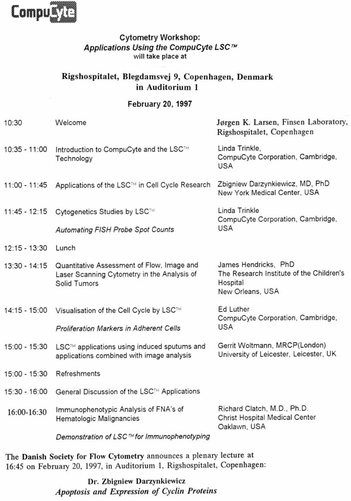


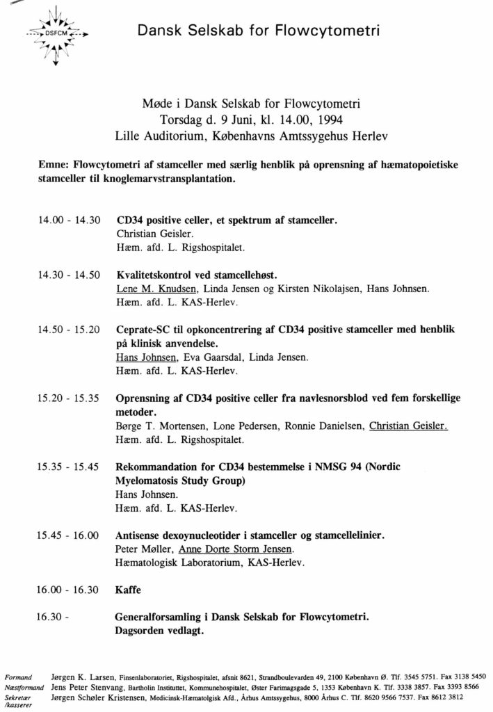

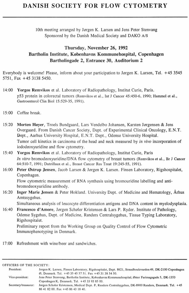
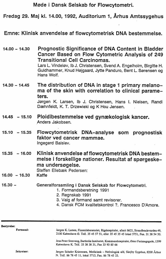
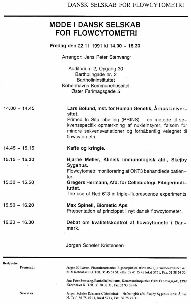
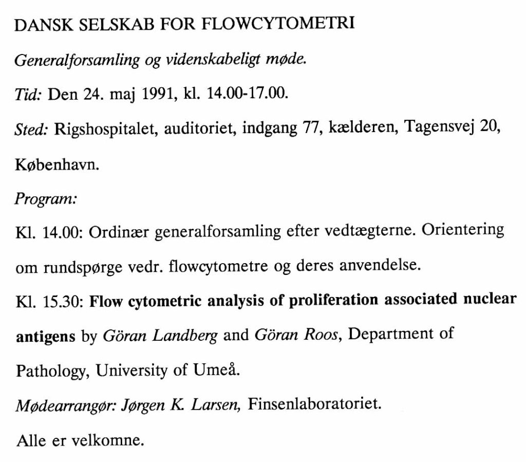


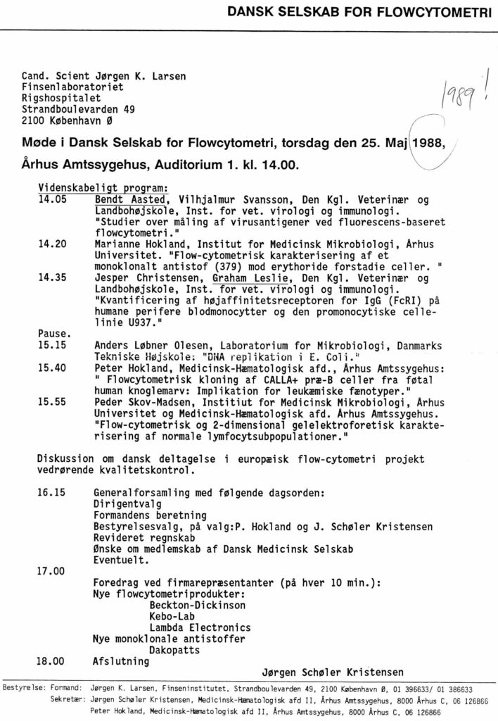

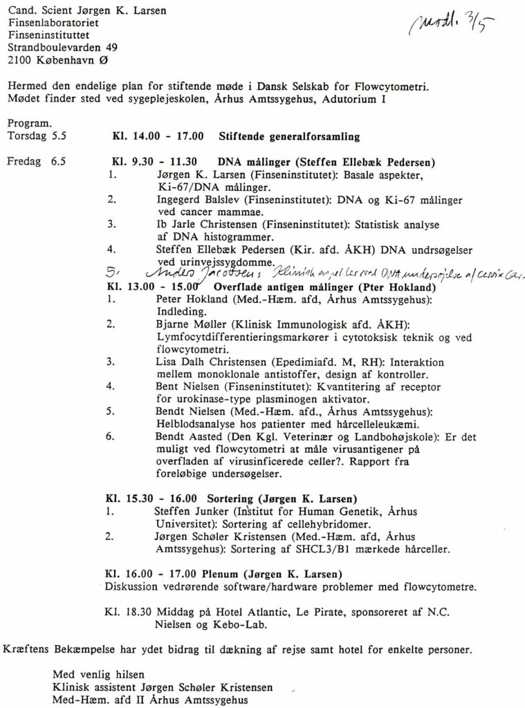